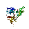
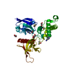
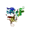
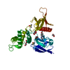
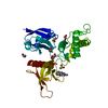
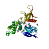
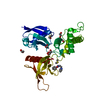
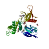
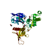
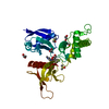
protein localization to paranode region of axon / cytoskeletal protein-membrane anchor activity / paranodal junction assembly / protein localization to juxtaparanode region of axon / myelin maintenance / paranode region of axon / cortical cytoskeleton organization / juxtaparanode region of axon / actomyosin structure organization / cortical actin cytoskeleton organization ...protein localization to paranode region of axon / cytoskeletal protein-membrane anchor activity / paranodal junction assembly / protein localization to juxtaparanode region of axon / myelin maintenance / paranode region of axon / cortical cytoskeleton organization / juxtaparanode region of axon / actomyosin structure organization / cortical actin cytoskeleton organization / Sensory processing of sound by outer hair cells of the cochlea / Neurexins and neuroligins / Sensory processing of sound by inner hair cells of the cochlea / regulation of neurotransmitter receptor localization to postsynaptic specialization membrane / neuron projection morphogenesis / protein localization to plasma membrane / structural constituent of cytoskeleton / cell-cell junction / cell junction / regulation of cell shape / actin binding / cytoskeleton / postsynapse / cilium / ciliary basal body / apoptotic process / glutamatergic synapse / plasma membrane / cytosol Similarity search - Function
Band 4.1-like protein 3 / SAB domain / Band 4.1, C-terminal / SAB domain / 4.1 protein C-terminal domain (CTD) / FERM adjacent (FA) / FERM adjacent (FA) / FERM adjacent (FA) / Acyl-CoA Binding Protein - #10 / Acyl-CoA Binding Protein ...Band 4.1-like protein 3 / SAB domain / Band 4.1, C-terminal / SAB domain / 4.1 protein C-terminal domain (CTD) / FERM adjacent (FA) / FERM adjacent (FA) / FERM adjacent (FA) / Acyl-CoA Binding Protein - #10 / Acyl-CoA Binding Protein / Ezrin/radixin/moesin-like / FERM, C-terminal PH-like domain / FERM C-terminal PH-like domain / FERM C-terminal PH-like domain / FERM, N-terminal / FERM N-terminal domain / FERM domain signature 1. / FERM conserved site / FERM domain signature 2. / FERM central domain / FERM/acyl-CoA-binding protein superfamily / Pleckstrin-homology domain (PH domain)/Phosphotyrosine-binding domain (PTB) / PH-domain like / FERM central domain / FERM superfamily, second domain / FERM domain / FERM domain profile. / Band 4.1 domain / Band 4.1 homologues / Phosphatidylinositol 3-kinase Catalytic Subunit; Chain A, domain 1 / Ubiquitin-like (UB roll) / PH-like domain superfamily / Roll / Ubiquitin-like domain superfamily / Roll / Up-down Bundle / Mainly Beta / Mainly Alpha / Alpha Beta Similarity search - Domain/homology
Resolution: 1.45→61.59 Å / Cor.coef. Fo:Fc: 0.97 / Cor.coef. Fo:Fc free: 0.952 / SU B: 2.926 / SU ML: 0.049 / Cross valid method: THROUGHOUT / ESU R: 0.07 / ESU R Free: 0.067 / Details: HYDROGENS HAVE BEEN ADDED IN THE RIDING POSITIONS
Rfactor
Num. reflection
% reflection
Selection details
Rfree
0.19994
1984
3.6 %
RANDOM
Rwork
0.15393
-
-
-
obs
0.15562
52770
99.99 %
-
Solvent computation
Ion probe radii: 0.8 Å / Shrinkage radii: 0.8 Å / VDW probe radii: 1.2 Å
 Movie
Movie Controller


 Open data
 Basic information
 Components
 Keywords
 Function and homology information
 Homo sapiens (human)
 X-RAY DIFFRACTION /
  Authors
 United States, 1items
  Citation
 Structure visualization
 Downloads & links
 Other downloads
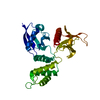


 PDBj
PDBj


 Assembly
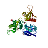


 Mass: 62.068 Da / Num. of mol.: 6 / Source method: obtained synthetically / Formula: C2H6O2
 Mass: 195.237 Da / Num. of mol.: 1 / Source method: obtained synthetically / Formula: C6H13NO4S / Comment: pH buffer*YM
 Mass: 18.015 Da / Num. of mol.: 183 / Source method: isolated from a natural source / Formula: H2O
 Sample preparation
 / Beamline: I03 / Wavelength: 0.9762 Å
 Processing