 Movie
Movie Controller


+ Open data

- Basic information
| Entry | Database: PDB / ID: 1c0n | ||||||
|---|---|---|---|---|---|---|---|
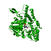
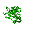
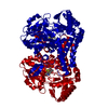
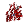
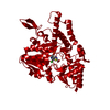
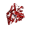
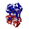
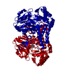
| Title | CSDB PROTEIN, NIFS HOMOLOGUE | ||||||
 Components Components | PROTEIN (CSDB PROTEIN) | ||||||
 Keywords Keywords | LYASE / ALPHA/BETA FOLD | ||||||
| Function / homology |  Function and homology information Function and homology informationselenium compound metabolic process / Hydrolases; Acting on carbon-sulfur bonds; Acting on carbon-sulfur bonds / cysteine sulfinate desulfinase activity / selenocysteine lyase / selenocysteine lyase activity / sulfur compound metabolic process / cysteine desulfurase / cysteine desulfurase activity / : / iron-sulfur cluster assembly ...selenium compound metabolic process / Hydrolases; Acting on carbon-sulfur bonds; Acting on carbon-sulfur bonds / cysteine sulfinate desulfinase activity / selenocysteine lyase / selenocysteine lyase activity / sulfur compound metabolic process / cysteine desulfurase / cysteine desulfurase activity / : / iron-sulfur cluster assembly / pyridoxal phosphate binding / hydrolase activity / protein homodimerization activity / cytoplasm Similarity search - Function | ||||||
| Biological species |  | ||||||
| Method |  X-RAY DIFFRACTION / Resolution: 2.8 Å X-RAY DIFFRACTION / Resolution: 2.8 Å | ||||||
 Authors Authors | Fujii, T. / Maeda, M. / Mihara, H. / Kurihara, T. / Esaki, N. / Hata, Y. | ||||||
 Citation Citation | Journal: Biochemistry / Year: 2000 Title: Structure of a NifS homologue: X-ray structure analysis of CsdB, an Escherichia coli counterpart of mammalian selenocysteine lyase Authors: Fujii, T. / Maeda, M. / Mihara, H. / Kurihara, T. / Esaki, N. / Hata, Y. #1: Journal: J.Biol.Chem. / Year: 1999Title: A nifS-like Gene, csdB, Encodes an Escherichia coli Counterpart of Mammalian Selenocysteine Lyase. GENE CLONING, PURIFICATION, CHARACTERIZATION AND PRELIMINARY X-RAY CRYSTALLOGRAPHIC STUDIES Authors: Mihara, H. / Maeda, M. / Fujii, T. / Kurihara, T. / Hata, Y. / Esaki, N. | ||||||
| History |
|
- Structure visualization
| Structure viewer | Molecule: MolmilJmol/JSmol |
|---|
- Downloads & links
-Download
| PDBx/mmCIF format | 1c0n.cif.gz | 87.6 KB | Display | PDBx/mmCIF format |
|---|---|---|---|---|
| PDB format | pdb1c0n.ent.gz | 67.4 KB | Display | PDB format |
| PDBx/mmJSON format | 1c0n.json.gz | Tree view | PDBx/mmJSON format | |
| Others |  Other downloads Other downloads |
-Validation report
| Arichive directory | https://data.pdbj.org/pub/pdb/validation_reports/c0/1c0nftp://data.pdbj.org/pub/pdb/validation_reports/c0/1c0n | HTTPS FTP |
|---|
-Related structure data
| Similar structure data |
|---|


-Links
 PDBj
PDBj

- Assembly
| Deposited unit | 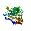
| ||||||||
|---|---|---|---|---|---|---|---|---|---|
| 1 | 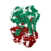
| ||||||||
| Unit cell |
|
-Components
| #1: Protein | Mass: 44077.035 Da / Num. of mol.: 1 Source method: isolated from a genetically manipulated source Source: (gene. exp.) |
|---|---|
| #2: Chemical | ChemComp-PLP /   Mass: 247.142 Da / Num. of mol.: 1 / Source method: obtained synthetically / Formula: C8H10NO6P Mass: 247.142 Da / Num. of mol.: 1 / Source method: obtained synthetically / Formula: C8H10NO6P |
| #3: Chemical | ChemComp-ACY /   Mass: 60.052 Da / Num. of mol.: 1 / Source method: obtained synthetically / Formula: C2H4O2 Mass: 60.052 Da / Num. of mol.: 1 / Source method: obtained synthetically / Formula: C2H4O2 |
| Has protein modification | N |
-Experimental details
-Experiment
| Experiment | Method: X-RAY DIFFRACTION / Number of used crystals: 1 |
|---|
- Sample preparation
| Crystal grow | Temperature: 298 K / Method: vapor diffusion, hanging drop / pH: 6.8 Details: sodium acetate, pottasium phosphate, pH 6.8, VAPOR DIFFUSION, HANGING DROP, temperature 298.0K | |||||||||||||||||||||||||
|---|---|---|---|---|---|---|---|---|---|---|---|---|---|---|---|---|---|---|---|---|---|---|---|---|---|---|
| Crystal | *PLUS Density % sol: 62 % | |||||||||||||||||||||||||
| Crystal grow | *PLUS Temperature: 25 ℃ | |||||||||||||||||||||||||
| Components of the solutions | *PLUS
|
-Data collection
| Diffraction | Mean temperature: 293 K |
|---|---|
| Diffraction source | Source: ROTATING ANODE / Type: RIGAKU RU300 / Wavelength: 1.5418 |
| Detector | Type: RIGAKU RAXIS IIC / Detector: IMAGE PLATE / Date: Jun 17, 1997 |
| Radiation | Protocol: SINGLE WAVELENGTH / Monochromatic (M) / Laue (L): M / Scattering type: x-ray |
| Radiation wavelength | Wavelength: 1.5418 Å / Relative weight: 1 |
| Reflection | Resolution: 2.8→93.6 Å / Num. all: 27061 / Num. obs: 27061 / % possible obs: 94.2 % / Observed criterion σ(I): 1 / Redundancy: 2.7 % / Biso Wilson estimate: 44.1 Å2 / Rmerge(I) obs: 0.072 / Net I/σ(I): 9.62 |
| Reflection shell | Resolution: 2.8→3 Å / Rmerge(I) obs: 0.216 / Num. unique all: 4641 / % possible all: 88.5 |
| Reflection | *PLUS Num. obs: 23770 / Num. measured all: 73138 |
| Reflection shell | *PLUS % possible obs: 88.5 % |
- Processing
| Software |
| |||||||||||||||
|---|---|---|---|---|---|---|---|---|---|---|---|---|---|---|---|---|
| Refinement | Resolution: 2.8→8 Å / σ(F): 1 / Stereochemistry target values: Engh & Huber
| |||||||||||||||
| Refinement step | Cycle: LAST / Resolution: 2.8→8 Å
| |||||||||||||||
| Refine LS restraints |
| |||||||||||||||
| Software | *PLUS Name: X-PLOR / Version: 3.851 / Classification: refinement | |||||||||||||||
| LS refinement shell | *PLUS Highest resolution: 2.8 Å / Lowest resolution: 2.92 Å / Rfactor Rfree: 0.345 / Rfactor Rwork: 0.29 / Num. reflection obs: 2000 |
