 Movie
Movie Controller


+ Open data

- Basic information
| Entry | Database: PDB / ID: 4rnj | ||||||
|---|---|---|---|---|---|---|---|
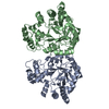
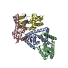
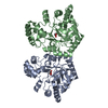
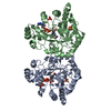
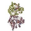
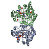
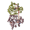
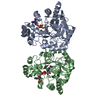
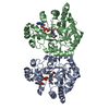
| Title | PaMorA phosphodiesterase domain, apo form | ||||||
 Components Components | Motility regulator | ||||||
 Keywords Keywords | HYDROLASE / EAL domain / Phosphodiesterase / c-di-GMP | ||||||
| Function / homology |  Function and homology information Function and homology informationc-di-GMP signaling / cyclic-guanylate-specific phosphodiesterase / cyclic-guanylate-specific phosphodiesterase activity / diguanylate cyclase activity / cellular response to nitric oxide / nucleotide binding / regulation of DNA-templated transcription / metal ion binding / identical protein binding / plasma membrane Similarity search - Function | ||||||
| Biological species |  Pseudomonas aeruginosa PAO1 (bacteria) Pseudomonas aeruginosa PAO1 (bacteria) | ||||||
| Method |  X-RAY DIFFRACTION / SYNCHROTRON / MOLECULAR REPLACEMENT / Resolution: 2.32 Å X-RAY DIFFRACTION / SYNCHROTRON / MOLECULAR REPLACEMENT / Resolution: 2.32 Å | ||||||
 Authors Authors | Phippen, C.W. / Tews, I. | ||||||
 Citation Citation | Journal: Febs Lett. / Year: 2014 Title: Formation and dimerization of the phosphodiesterase active site of the Pseudomonas aeruginosa MorA, a bi-functional c-di-GMP regulator. Authors: Phippen, C.W. / Mikolajek, H. / Schlaefli, H.G. / Keevil, C.W. / Webb, J.S. / Tews, I. | ||||||
| History |
|
- Structure visualization
| Structure viewer | Molecule: MolmilJmol/JSmol |
|---|
- Downloads & links
-Download
| PDBx/mmCIF format | 4rnj.cif.gz | 113.7 KB | Display | PDBx/mmCIF format |
|---|---|---|---|---|
| PDB format | pdb4rnj.ent.gz | 86.1 KB | Display | PDB format |
| PDBx/mmJSON format | 4rnj.json.gz | Tree view | PDBx/mmJSON format | |
| Others |  Other downloads Other downloads |
-Validation report
| Arichive directory | https://data.pdbj.org/pub/pdb/validation_reports/rn/4rnjftp://data.pdbj.org/pub/pdb/validation_reports/rn/4rnj | HTTPS FTP |
|---|
-Related structure data
| Related structure data |  4rnfC 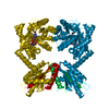 4rnhC  4rniC  3n3tS C: citing same article ( S: Starting model for refinement |
|---|---|
| Similar structure data |

-Links
 PDBj
PDBj


- Assembly
| Deposited unit | 
| ||||||||
|---|---|---|---|---|---|---|---|---|---|
| 1 |
| ||||||||
| 2 | 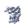
| ||||||||
| 3 | 
| ||||||||
| Unit cell |
| ||||||||
| Details | Active diguanylate cyclase |
-Components
| #1: Protein | Mass: 31727.320 Da / Num. of mol.: 2 / Fragment: EAL domain, UNP residues 1145-1409 Source method: isolated from a genetically manipulated source Source: (gene. exp.) Pseudomonas aeruginosa PAO1 (bacteria) / Strain: PA01 / Gene: morA, PA4601 / Plasmid: pET28a / Production host: References: UniProt: Q9HVI8, cyclic-guanylate-specific phosphodiesterase #2: Water | ChemComp-HOH / |  Mass: 18.015 Da / Num. of mol.: 117 / Source method: isolated from a natural source / Formula: H2O Mass: 18.015 Da / Num. of mol.: 117 / Source method: isolated from a natural source / Formula: H2O |
|---|
-Experimental details
-Experiment
| Experiment | Method: X-RAY DIFFRACTION / Number of used crystals: 1 |
|---|
- Sample preparation
| Crystal | Density Matthews: 2.27 Å3/Da / Density % sol: 45.82 % |
|---|---|
| Crystal grow | Temperature: 295 K / Method: vapor diffusion, sitting drop / pH: 6.5 Details: 0.1M Imidazole; MES, 0.09M NaN03; Na2HPO4; (NH4)2SO4, 30% Ethylene glycol; PEG 8000, pH 6.5, VAPOR DIFFUSION, SITTING DROP, temperature 295K |
-Data collection
| Diffraction | Mean temperature: 100 K |
|---|---|
| Diffraction source | Source: SYNCHROTRON / Site: ESRF  / Beamline: ID14-1 / Wavelength: 0.934 Å / Beamline: ID14-1 / Wavelength: 0.934 Å |
| Detector | Type: ADSC QUANTUM 210 / Detector: CCD / Date: Sep 14, 2012 |
| Radiation | Monochromator: Diamond (001) / Protocol: SINGLE WAVELENGTH / Monochromatic (M) / Laue (L): M / Scattering type: x-ray |
| Radiation wavelength | Wavelength: 0.934 Å / Relative weight: 1 |
| Reflection | Resolution: 2.32→100 Å / Num. all: 25794 / Num. obs: 25720 / % possible obs: 99.7 % / Redundancy: 4.28 % / Biso Wilson estimate: 42.67 Å2 / Rmerge(I) obs: 0.072 / Net I/σ(I): 13.87 |
| Reflection shell | Resolution: 2.32→2.4 Å / Redundancy: 3.69 % / Rmerge(I) obs: 0.678 / Mean I/σ(I) obs: 1.8 / % possible all: 98.4 |
- Processing
| Software |
| ||||||||||||||||||||||||||||||||||||||||||||||||||||||||||||||||||||||
|---|---|---|---|---|---|---|---|---|---|---|---|---|---|---|---|---|---|---|---|---|---|---|---|---|---|---|---|---|---|---|---|---|---|---|---|---|---|---|---|---|---|---|---|---|---|---|---|---|---|---|---|---|---|---|---|---|---|---|---|---|---|---|---|---|---|---|---|---|---|---|---|
| Refinement | Method to determine structure: MOLECULAR REPLACEMENT Starting model: PDB ENTRY 3N3T Resolution: 2.32→48.741 Å / FOM work R set: 0.8062 / SU ML: 0.28 / Cross valid method: THROUGHOUT / σ(F): 1.34 / Phase error: 25.77 / Stereochemistry target values: MAXIMUM LIKELIHOOD Details: HYDROGENS HAVE BEEN ADDED IN THE RIDING POSITIONS U VALUES : REFINED INDIVIDUALLY
| ||||||||||||||||||||||||||||||||||||||||||||||||||||||||||||||||||||||
| Solvent computation | Shrinkage radii: 0.9 Å / VDW probe radii: 1.11 Å / Solvent model: FLAT BULK SOLVENT MODEL | ||||||||||||||||||||||||||||||||||||||||||||||||||||||||||||||||||||||
| Displacement parameters | Biso max: 119.79 Å2 / Biso mean: 47.68 Å2 / Biso min: 25.23 Å2 | ||||||||||||||||||||||||||||||||||||||||||||||||||||||||||||||||||||||
| Refinement step | Cycle: LAST / Resolution: 2.32→48.741 Å
| ||||||||||||||||||||||||||||||||||||||||||||||||||||||||||||||||||||||
| Refine LS restraints |
| ||||||||||||||||||||||||||||||||||||||||||||||||||||||||||||||||||||||
| LS refinement shell | Refine-ID: X-RAY DIFFRACTION / Total num. of bins used: 9
|
