 Movie
Movie Controller

[English] 日本語
 Yorodumi
- PDB-4q0e: Crystal structure of TS-DHFR from Cryptosporidium hominis in comp... -
+ Open data

- Basic information
| Entry | Database: PDB / ID: 4q0e | ||||||
|---|---|---|---|---|---|---|---|
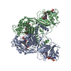
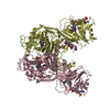
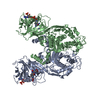
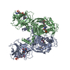
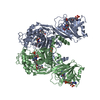
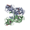
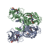
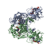
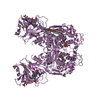
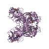
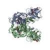
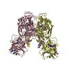
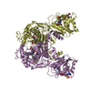
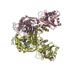
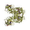
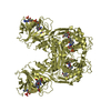
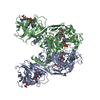
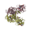
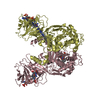
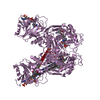
| Title | Crystal structure of TS-DHFR from Cryptosporidium hominis in complex with NADPH, FdUMP and 2-amino-4-oxo-4,7-dihydro-pyrrolo[2,3-d]pyrimidine-methyl-phenyl-L-glutamic acid. | ||||||
 Components Components | Bifunctional dihydrofolate reductase-thymidylate synthase | ||||||
 Keywords Keywords | transferase/transferase inhibitor / bifunctional enzyme / transferase / oxidoreductase / transferase-transferase inhibitor complex | ||||||
| Function / homology |  Function and homology information Function and homology informationThymidylate Synthase; Chain A / Thymidylate synthase/dCMP hydroxymethylase domain / Dihydrofolate Reductase, subunit A / Dihydrofolate Reductase, subunit A / 2-Layer Sandwich / 3-Layer(aba) Sandwich / Alpha Beta Similarity search - Domain/homology | ||||||
| Biological species |  Cryptosporidium hominis (eukaryote) Cryptosporidium hominis (eukaryote) | ||||||
| Method |  X-RAY DIFFRACTION / SYNCHROTRON / MOLECULAR REPLACEMENT / Resolution: 2.78 Å X-RAY DIFFRACTION / SYNCHROTRON / MOLECULAR REPLACEMENT / Resolution: 2.78 Å | ||||||
 Authors Authors | Kumar, V.P. / Anderson, K.S. | ||||||
 Citation Citation | Journal: Bioorg.Med.Chem.Lett. / Year: 2014 Title: Structural studies provide clues for analog design of specific inhibitors of Cryptosporidium hominis thymidylate synthase-dihydrofolate reductase. Authors: Kumar, V.P. / Cisneros, J.A. / Frey, K.M. / Castellanos-Gonzalez, A. / Wang, Y. / Gangjee, A. / White, A.C. / Jorgensen, W.L. / Anderson, K.S. | ||||||
| History |
|
- Structure visualization
| Structure viewer | Molecule: MolmilJmol/JSmol |
|---|
- Downloads & links
-Download
| PDBx/mmCIF format | 4q0e.cif.gz | 521 KB | Display | PDBx/mmCIF format |
|---|---|---|---|---|
| PDB format | pdb4q0e.ent.gz | 431.6 KB | Display | PDB format |
| PDBx/mmJSON format | 4q0e.json.gz | Tree view | PDBx/mmJSON format | |
| Others |  Other downloads Other downloads |
-Validation report
| Arichive directory | https://data.pdbj.org/pub/pdb/validation_reports/q0/4q0eftp://data.pdbj.org/pub/pdb/validation_reports/q0/4q0e | HTTPS FTP |
|---|
-Related structure data
| Related structure data |  4q0dC  4ky8S S: Starting model for refinement C: citing same article ( |
|---|---|
| Similar structure data |


-Links
 PDBj
PDBj

- Assembly
| Deposited unit | 
| |||||||||||||||||||||||||||||||||||||||||||||||||||||||||||||||||||||||||||||||||||||||||||||||||||
|---|---|---|---|---|---|---|---|---|---|---|---|---|---|---|---|---|---|---|---|---|---|---|---|---|---|---|---|---|---|---|---|---|---|---|---|---|---|---|---|---|---|---|---|---|---|---|---|---|---|---|---|---|---|---|---|---|---|---|---|---|---|---|---|---|---|---|---|---|---|---|---|---|---|---|---|---|---|---|---|---|---|---|---|---|---|---|---|---|---|---|---|---|---|---|---|---|---|---|---|---|
| 1 | 
| |||||||||||||||||||||||||||||||||||||||||||||||||||||||||||||||||||||||||||||||||||||||||||||||||||
| 2 | 
| |||||||||||||||||||||||||||||||||||||||||||||||||||||||||||||||||||||||||||||||||||||||||||||||||||
| 3 | 
| |||||||||||||||||||||||||||||||||||||||||||||||||||||||||||||||||||||||||||||||||||||||||||||||||||
| Unit cell |
| |||||||||||||||||||||||||||||||||||||||||||||||||||||||||||||||||||||||||||||||||||||||||||||||||||
| Noncrystallographic symmetry (NCS) | NCS domain:
NCS domain segments: Ens-ID: 1
|