 Movie
Movie Controller


+ Open data

- Basic information
| Entry | Database: PDB / ID: 1qzf | ||||||
|---|---|---|---|---|---|---|---|
| Title | Crystal structure of DHFR-TS from Cryptosporidium hominis | ||||||
 Components Components | bifunctional dihydrofolate reductase-thymidylate synthase | ||||||
 Keywords Keywords | OXIDOREDUCTASE / TRANSFERASE / bifunctional enzyme | ||||||
| Function / homology |  Function and homology information Function and homology informationthymidylate synthase activity / dTMP biosynthetic process / dihydrofolate reductase activity / tetrahydrofolate biosynthetic process / one-carbon metabolic process / methylation / nucleotide binding / mitochondrion / cytosol Similarity search - Function | ||||||
| Biological species |  Cryptosporidium hominis (eukaryote) Cryptosporidium hominis (eukaryote) | ||||||
| Method |  X-RAY DIFFRACTION / SYNCHROTRON / MOLECULAR REPLACEMENT / Resolution: 2.8 Å X-RAY DIFFRACTION / SYNCHROTRON / MOLECULAR REPLACEMENT / Resolution: 2.8 Å | ||||||
 Authors Authors | O'Neil, R.H. / Lilien, R.H. / Donald, B.R. / Stroud, R.M. / Anderson, A.C. | ||||||
 Citation Citation | Journal: J.Biol.Chem. / Year: 2003 Title: Phylogenetic classification of protozoa based on the structure of the linker domain in the bifunctional enzyme, dihydrofolate reductase-thymidylate synthase Authors: O'Neil, R.H. / Lilien, R.H. / Donald, B.R. / Stroud, R.M. / Anderson, A.C. | ||||||
| History |
|
- Structure visualization
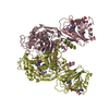
| Structure viewer | Molecule: MolmilJmol/JSmol |
|---|
- Downloads & links
-Download
| PDBx/mmCIF format | 1qzf.cif.gz | 541.9 KB | Display | PDBx/mmCIF format |
|---|---|---|---|---|
| PDB format | pdb1qzf.ent.gz | 445.5 KB | Display | PDB format |
| PDBx/mmJSON format | 1qzf.json.gz | Tree view | PDBx/mmJSON format | |
| Others |  Other downloads Other downloads |
-Validation report
| Arichive directory | https://data.pdbj.org/pub/pdb/validation_reports/qz/1qzfftp://data.pdbj.org/pub/pdb/validation_reports/qz/1qzf | HTTPS FTP |
|---|
-Related structure data


-Links
 PDBj
PDBj

- Assembly
| Deposited unit | 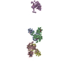
| ||||||||
|---|---|---|---|---|---|---|---|---|---|
| 1 | 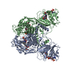
| ||||||||
| 2 | 
| ||||||||
| 3 | 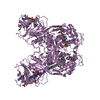
| ||||||||
| Unit cell |
| ||||||||
| Components on special symmetry positions |
| ||||||||
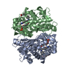
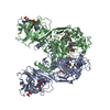
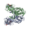
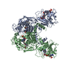
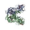
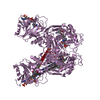
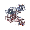
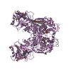
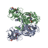
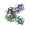
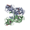
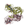
| Details | The biological assembly is a dimer. Two dimers (chains A,B and C,D) are present in the asymmetric unit and another dimer is generated using the monomer, E, and the two-fold axis: -x,y,-z |
-Components
-Protein , 1 types, 5 molecules ABCDE
| #1: Protein | Mass: 60262.520 Da / Num. of mol.: 5 Source method: isolated from a genetically manipulated source Source: (gene. exp.) Cryptosporidium hominis (eukaryote) / Production host:  References: UniProt: Q27552, dihydrofolate reductase, thymidylate synthase |
|---|
-Non-polymers , 5 types, 426 molecules 
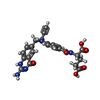
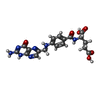


| #2: Chemical | ChemComp-UMP /  Mass: 308.182 Da / Num. of mol.: 5 / Source method: obtained synthetically / Formula: C9H13N2O8P Mass: 308.182 Da / Num. of mol.: 5 / Source method: obtained synthetically / Formula: C9H13N2O8P#3: Chemical | ChemComp-CB3 /  Mass: 477.469 Da / Num. of mol.: 5 / Source method: obtained synthetically / Formula: C24H23N5O6 Mass: 477.469 Da / Num. of mol.: 5 / Source method: obtained synthetically / Formula: C24H23N5O6#4: Chemical | ChemComp-FOL /  Mass: 441.397 Da / Num. of mol.: 5 / Source method: obtained synthetically / Formula: C19H19N7O6 Mass: 441.397 Da / Num. of mol.: 5 / Source method: obtained synthetically / Formula: C19H19N7O6#5: Chemical | ChemComp-NDP /  Mass: 745.421 Da / Num. of mol.: 5 / Source method: obtained synthetically / Formula: C21H30N7O17P3 Mass: 745.421 Da / Num. of mol.: 5 / Source method: obtained synthetically / Formula: C21H30N7O17P3#6: Water | ChemComp-HOH / | Mass: 18.015 Da / Num. of mol.: 406 / Source method: isolated from a natural source / Formula: H2O |
|---|
-Experimental details
-Experiment
| Experiment | Method: X-RAY DIFFRACTION / Number of used crystals: 1 |
|---|
- Sample preparation
| Crystal | Density Matthews: 4.54 Å3/Da / Density % sol: 72.88 % | ||||||||||||||||||||||||||||||||||||
|---|---|---|---|---|---|---|---|---|---|---|---|---|---|---|---|---|---|---|---|---|---|---|---|---|---|---|---|---|---|---|---|---|---|---|---|---|---|
| Crystal grow | Temperature: 298 K / Method: vapor diffusion, hanging drop / pH: 8 Details: PEG 6000, ammonium sulfate, lithium sulfate, Tris, pH 8.0, VAPOR DIFFUSION, HANGING DROP, temperature 298K | ||||||||||||||||||||||||||||||||||||
| Crystal grow | *PLUS Method: vapor diffusion, hanging drop | ||||||||||||||||||||||||||||||||||||
| Components of the solutions | *PLUS
|
-Data collection
| Diffraction | Mean temperature: 100 K |
|---|---|
| Diffraction source | Source: SYNCHROTRON / Site: NSLS  / Beamline: X12C / Wavelength: 1.08 Å / Beamline: X12C / Wavelength: 1.08 Å |
| Detector | Type: BRANDEIS - B4 / Detector: CCD / Date: Apr 5, 1998 |
| Radiation | Monochromator: Si 111 channel / Protocol: SINGLE WAVELENGTH / Monochromatic (M) / Laue (L): M / Scattering type: x-ray |
| Radiation wavelength | Wavelength: 1.08 Å / Relative weight: 1 |
| Reflection | Resolution: 2.8→30 Å / Num. obs: 134842 / % possible obs: 87.9 % / Observed criterion σ(F): 2 / Observed criterion σ(I): 2 / Rmerge(I) obs: 0.105 / Net I/σ(I): 8.7 |
| Reflection shell | Resolution: 2.8→2.9 Å / % possible all: 52.1 |
- Processing
| Software |
| ||||||||||||||||||||||||||||||||||||
|---|---|---|---|---|---|---|---|---|---|---|---|---|---|---|---|---|---|---|---|---|---|---|---|---|---|---|---|---|---|---|---|---|---|---|---|---|---|
| Refinement | Method to determine structure: MOLECULAR REPLACEMENT Starting model: PDB ENTRIES 1f28 AND 1cd2 Resolution: 2.8→29.8 Å / Rfactor Rfree error: 0.003 / Data cutoff high absF: 3828805.98 / Data cutoff high rms absF: 3828805.98 / Data cutoff low absF: 0 / Isotropic thermal model: RESTRAINED / Cross valid method: THROUGHOUT / σ(F): 0 / Stereochemistry target values: Engh & Huber
| ||||||||||||||||||||||||||||||||||||
| Solvent computation | Solvent model: FLAT MODEL / Bsol: 21.4533 Å2 / ksol: 0.324071 e/Å3 | ||||||||||||||||||||||||||||||||||||
| Displacement parameters | Biso mean: 38.9 Å2
| ||||||||||||||||||||||||||||||||||||
| Refine analyze |
| ||||||||||||||||||||||||||||||||||||
| Refinement step | Cycle: LAST / Resolution: 2.8→29.8 Å
| ||||||||||||||||||||||||||||||||||||
| Refine LS restraints |
| ||||||||||||||||||||||||||||||||||||
| LS refinement shell | Resolution: 2.8→2.98 Å / Rfactor Rfree error: 0.015 / Total num. of bins used: 6
| ||||||||||||||||||||||||||||||||||||
| Xplor file |
| ||||||||||||||||||||||||||||||||||||
| Refinement | *PLUS Highest resolution: 2.8 Å | ||||||||||||||||||||||||||||||||||||
| Solvent computation | *PLUS | ||||||||||||||||||||||||||||||||||||
| Displacement parameters | *PLUS | ||||||||||||||||||||||||||||||||||||
| Refine LS restraints | *PLUS
|
