 Movie
Movie Controller

[English] 日本語
 Yorodumi
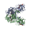
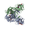
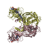
- PDB-2oip: Crystal Structure of the S290G Active Site Mutant of TS-DHFR from... -
+ Open data

- Basic information
| Entry | Database: PDB / ID: 2oip | ||||||
|---|---|---|---|---|---|---|---|
| Title | Crystal Structure of the S290G Active Site Mutant of TS-DHFR from Cryptosporidium hominis | ||||||
 Components Components | Chain A, crystal structure of Dhfr | ||||||
 Keywords Keywords | TRANSFERASE / OXIDOREDUCTASE / BIFUNCTIONAL ENZYME | ||||||
| Function / homology |  Function and homology information Function and homology informationThymidylate Synthase; Chain A / Thymidylate synthase/dCMP hydroxymethylase domain / Dihydrofolate Reductase, subunit A / Dihydrofolate Reductase, subunit A / 2-Layer Sandwich / 3-Layer(aba) Sandwich / Alpha Beta Similarity search - Domain/homology | ||||||
| Biological species |  Cryptosporidium hominis (eukaryote) Cryptosporidium hominis (eukaryote) | ||||||
| Method |  X-RAY DIFFRACTION / SYNCHROTRON / MOLECULAR REPLACEMENT / Resolution: 2.8 Å X-RAY DIFFRACTION / SYNCHROTRON / MOLECULAR REPLACEMENT / Resolution: 2.8 Å | ||||||
 Authors Authors | Martucci, W.E. / Vargo, M.A. | ||||||
 Citation Citation | Journal: Biochemistry / Year: 2007 Title: Nonconserved residues Ala287 and Ser290 of the Cryptosporidium hominis thymidylate synthase domain facilitate its rapid rate of catalysis Authors: Doan, L.T. / Martucci, W.E. / Vargo, M.A. / Atreya, C.E. / Anderson, K.S. | ||||||
| History |
|
- Structure visualization
| Structure viewer | Molecule: MolmilJmol/JSmol |
|---|
- Downloads & links
-Download
| PDBx/mmCIF format | 2oip.cif.gz | 539.9 KB | Display | PDBx/mmCIF format |
|---|---|---|---|---|
| PDB format | pdb2oip.ent.gz | 445.3 KB | Display | PDB format |
| PDBx/mmJSON format | 2oip.json.gz | Tree view | PDBx/mmJSON format | |
| Others |  Other downloads Other downloads |
-Validation report
| Arichive directory | https://data.pdbj.org/pub/pdb/validation_reports/oi/2oipftp://data.pdbj.org/pub/pdb/validation_reports/oi/2oip | HTTPS FTP |
|---|
-Related structure data
| Related structure data |  1qzfS S: Starting model for refinement |
|---|---|
| Similar structure data |


-Links
 PDBj
PDBj

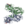
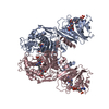
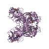
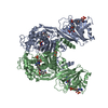
- Assembly
| Deposited unit | 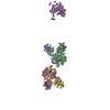
| ||||||||
|---|---|---|---|---|---|---|---|---|---|
| 1 | 
| ||||||||
| 2 | 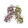
| ||||||||
| 3 | 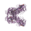
| ||||||||
| Unit cell |
| ||||||||
| Details | The biological assembly is the dimer present in the asymmetric unit, either with monomers A+B or C+D. The second half of the dimer formed by monomer E lies across the crystallographic axis. |
-Components
-Protein , 1 types, 5 molecules ABCDE
| #1: Protein | Mass: 60014.219 Da / Num. of mol.: 5 / Mutation: S290G Source method: isolated from a genetically manipulated source Source: (gene. exp.) Cryptosporidium hominis (eukaryote) / Species (production host): Escherichia coli / Production host:  References: UniProt: Q5CGA3, thymidylate synthase, dihydrofolate reductase |
|---|
-Non-polymers , 5 types, 424 molecules 
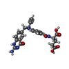
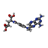


| #2: Chemical | ChemComp-UMP /  Mass: 308.182 Da / Num. of mol.: 5 / Source method: obtained synthetically / Formula: C9H13N2O8P Mass: 308.182 Da / Num. of mol.: 5 / Source method: obtained synthetically / Formula: C9H13N2O8P#3: Chemical | ChemComp-CB3 /  Mass: 477.469 Da / Num. of mol.: 5 / Source method: obtained synthetically / Formula: C24H23N5O6 Mass: 477.469 Da / Num. of mol.: 5 / Source method: obtained synthetically / Formula: C24H23N5O6#4: Chemical | ChemComp-MTX /  Mass: 454.439 Da / Num. of mol.: 5 / Source method: obtained synthetically / Formula: C20H22N8O5 Mass: 454.439 Da / Num. of mol.: 5 / Source method: obtained synthetically / Formula: C20H22N8O5#5: Chemical | ChemComp-NDP /  Mass: 745.421 Da / Num. of mol.: 5 / Source method: obtained synthetically / Formula: C21H30N7O17P3 Mass: 745.421 Da / Num. of mol.: 5 / Source method: obtained synthetically / Formula: C21H30N7O17P3#6: Water | ChemComp-HOH / | Mass: 18.015 Da / Num. of mol.: 404 / Source method: isolated from a natural source / Formula: H2O |
|---|
-Details
| Has protein modification | N |
|---|
-Experimental details
-Experiment
| Experiment | Method: X-RAY DIFFRACTION / Number of used crystals: 1 |
|---|
- Sample preparation
| Crystal | Density Matthews: 4.49 Å3/Da / Density % sol: 72.63 % |
|---|---|
| Crystal grow | Temperature: 291 K / Method: vapor diffusion, hanging drop / pH: 8 Details: 0.1mM ammonium sulfate, 0.3M lithium sulfate, 0.1M Tris, 10% PEG 6000, pH 8.0, VAPOR DIFFUSION, HANGING DROP, temperature 291K |
-Data collection
| Diffraction | Mean temperature: 100 K |
|---|---|
| Diffraction source | Source: SYNCHROTRON / Site: NSLS  / Beamline: X25 / Wavelength: 1.1 Å / Beamline: X25 / Wavelength: 1.1 Å |
| Detector | Type: ADSC QUANTUM 315 / Detector: CCD / Date: Sep 9, 2005 |
| Radiation | Monochromator: Si(111) / Protocol: SINGLE WAVELENGTH / Monochromatic (M) / Laue (L): M / Scattering type: x-ray |
| Radiation wavelength | Wavelength: 1.1 Å / Relative weight: 1 |
| Reflection | Resolution: 2.8→50 Å / Num. obs: 130177 / % possible obs: 99.5 % / Observed criterion σ(F): 2 / Observed criterion σ(I): 2 / Redundancy: 3.3 % / Biso Wilson estimate: 65 Å2 / Rmerge(I) obs: 0.117 / Χ2: 1.234 / Net I/σ(I): 13.4 |
| Reflection shell | Resolution: 2.8→2.9 Å / Redundancy: 3.3 % / Rmerge(I) obs: 0.538 / Mean I/σ(I) obs: 2.9 / Num. unique all: 12987 / Χ2: 1.252 / % possible all: 99.6 |
-Phasing
| Phasing dm | FOM : 0.783 / FOM centric: 0.676 / Reflection: 131011 / Reflection centric: 4636 | ||||||||||||||||||||||||||||||||||||||||||||||||||
|---|---|---|---|---|---|---|---|---|---|---|---|---|---|---|---|---|---|---|---|---|---|---|---|---|---|---|---|---|---|---|---|---|---|---|---|---|---|---|---|---|---|---|---|---|---|---|---|---|---|---|---|
| Phasing dm shell |
|
- Processing
| Software |
| ||||||||||||||||||||||||||||||||||||
|---|---|---|---|---|---|---|---|---|---|---|---|---|---|---|---|---|---|---|---|---|---|---|---|---|---|---|---|---|---|---|---|---|---|---|---|---|---|
| Refinement | Method to determine structure: MOLECULAR REPLACEMENT Starting model: PDB Entry 1QZF Resolution: 2.8→50 Å / FOM work R set: 0.78 / Isotropic thermal model: group / Cross valid method: THROUGHOUT / σ(F): 0 / σ(I): 0 / Stereochemistry target values: Engh & Huber
| ||||||||||||||||||||||||||||||||||||
| Displacement parameters | Biso mean: 66.525 Å2
| ||||||||||||||||||||||||||||||||||||
| Refine analyze |
| ||||||||||||||||||||||||||||||||||||
| Refinement step | Cycle: LAST / Resolution: 2.8→50 Å
| ||||||||||||||||||||||||||||||||||||
| Refine LS restraints |
| ||||||||||||||||||||||||||||||||||||
| LS refinement shell | Resolution: 2.8→2.98 Å / Rfactor Rfree error: 0.012
|
