 Movie
Movie Controller

[English] 日本語
 Yorodumi
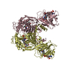
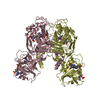
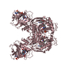
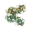
- PDB-3dl5: Crystal Structure of the A287F Active Site Mutant of TS-DHFR from... -
+ Open data

- Basic information
| Entry | Database: PDB / ID: 3dl5 | ||||||
|---|---|---|---|---|---|---|---|
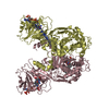
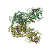
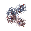
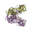
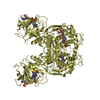
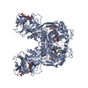
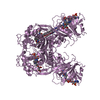
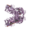
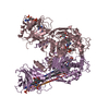
| Title | Crystal Structure of the A287F Active Site Mutant of TS-DHFR from Cryptosporidium hominis | ||||||
 Components Components | Dihydrofolate reductase, DHFR | ||||||
 Keywords Keywords | OXIDOREDUCTASE / Enzyme active site mutant / Enzyme-ligand complex | ||||||
| Function / homology |  Function and homology information Function and homology informationThymidylate Synthase; Chain A / Thymidylate synthase/dCMP hydroxymethylase domain / Dihydrofolate Reductase, subunit A / Dihydrofolate Reductase, subunit A / 2-Layer Sandwich / 3-Layer(aba) Sandwich / Alpha Beta Similarity search - Domain/homology | ||||||
| Biological species |  Cryptosporidium hominis (eukaryote) Cryptosporidium hominis (eukaryote) | ||||||
| Method |  X-RAY DIFFRACTION / SYNCHROTRON / MOLECULAR REPLACEMENT / Resolution: 2.74 Å X-RAY DIFFRACTION / SYNCHROTRON / MOLECULAR REPLACEMENT / Resolution: 2.74 Å | ||||||
 Authors Authors | Vargo, M.A. / Martucci, W.E. / Anderson, K.S. | ||||||
 Citation Citation | Journal: Biochemistry / Year: 2008 Title: Explaining an unusually fast parasitic enzyme: folate tail-binding residues dictate substrate positioning and catalysis in Cryptosporidium hominis thymidylate synthase. Authors: Martucci, W.E. / Vargo, M.A. / Anderson, K.S. | ||||||
| History |
|
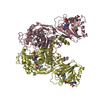
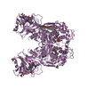
- Structure visualization
| Structure viewer | Molecule: MolmilJmol/JSmol |
|---|
- Downloads & links
-Download
| PDBx/mmCIF format | 3dl5.cif.gz | 536.3 KB | Display | PDBx/mmCIF format |
|---|---|---|---|---|
| PDB format | pdb3dl5.ent.gz | 442.4 KB | Display | PDB format |
| PDBx/mmJSON format | 3dl5.json.gz | Tree view | PDBx/mmJSON format | |
| Others |  Other downloads Other downloads |
-Validation report
| Arichive directory | https://data.pdbj.org/pub/pdb/validation_reports/dl/3dl5ftp://data.pdbj.org/pub/pdb/validation_reports/dl/3dl5 | HTTPS FTP |
|---|
-Related structure data
| Related structure data | 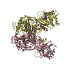 3dl6C 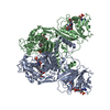 1qzfS C: citing same article ( S: Starting model for refinement |
|---|---|
| Similar structure data |


-Links
 PDBj
PDBj

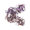
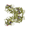
- Assembly
| Deposited unit | 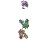
| ||||||||
|---|---|---|---|---|---|---|---|---|---|
| 1 | 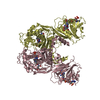
| ||||||||
| 2 | 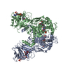
| ||||||||
| 3 | 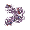
| ||||||||
| Unit cell |
|
-Components
-Protein , 1 types, 5 molecules ABCDE
| #1: Protein | Mass: 60338.613 Da / Num. of mol.: 5 / Mutation: A287F Source method: isolated from a genetically manipulated source Source: (gene. exp.) Cryptosporidium hominis (eukaryote) / Gene: Chro.40506 / Production host:  |
|---|
-Non-polymers , 5 types, 465 molecules 
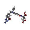


| #2: Chemical | ChemComp-UMP /  Mass: 308.182 Da / Num. of mol.: 5 / Source method: obtained synthetically / Formula: C9H13N2O8P Mass: 308.182 Da / Num. of mol.: 5 / Source method: obtained synthetically / Formula: C9H13N2O8P#3: Chemical | ChemComp-CB3 /  Mass: 477.469 Da / Num. of mol.: 5 / Source method: obtained synthetically / Formula: C24H23N5O6 Mass: 477.469 Da / Num. of mol.: 5 / Source method: obtained synthetically / Formula: C24H23N5O6#4: Chemical | ChemComp-DHF /  Mass: 443.413 Da / Num. of mol.: 5 / Source method: obtained synthetically / Formula: C19H21N7O6 Mass: 443.413 Da / Num. of mol.: 5 / Source method: obtained synthetically / Formula: C19H21N7O6#5: Chemical | ChemComp-NDP /  Mass: 745.421 Da / Num. of mol.: 5 / Source method: obtained synthetically / Formula: C21H30N7O17P3 Mass: 745.421 Da / Num. of mol.: 5 / Source method: obtained synthetically / Formula: C21H30N7O17P3#6: Water | ChemComp-HOH / | Mass: 18.015 Da / Num. of mol.: 445 / Source method: isolated from a natural source / Formula: H2O |
|---|
-Experimental details
-Experiment
| Experiment | Method: X-RAY DIFFRACTION / Number of used crystals: 1 |
|---|
- Sample preparation
| Crystal | Density Matthews: 4.55 Å3/Da / Density % sol: 72.94 % |
|---|---|
| Crystal grow | Temperature: 298 K / Method: vapor diffusion, hanging drop / pH: 8 Details: 0.1mM ammonium sulfate, 0.2mM, lithium sulfate, 0.1mM Tris, 12% PEG-6000, flash frozen in 25% ethylene glycol, pH 8.0, VAPOR DIFFUSION, HANGING DROP, temperature 298K |
-Data collection
| Diffraction | Mean temperature: 100 K |
|---|---|
| Diffraction source | Source: SYNCHROTRON / Site: NSLS  / Beamline: X29A / Wavelength: 1.08 Å / Beamline: X29A / Wavelength: 1.08 Å |
| Detector | Type: ADSC QUANTUM 315 / Detector: CCD / Date: Aug 16, 2006 |
| Radiation | Monochromator: Si (111) / Protocol: SINGLE WAVELENGTH / Monochromatic (M) / Laue (L): M / Scattering type: x-ray |
| Radiation wavelength | Wavelength: 1.08 Å / Relative weight: 1 |
| Reflection | Resolution: 2.74→45.4 Å / Num. obs: 141384 / % possible obs: 99.8 % / Observed criterion σ(F): 0 / Observed criterion σ(I): 0 / Redundancy: 3.6 % / Biso Wilson estimate: 68 Å2 / Rmerge(I) obs: 0.123 / Net I/σ(I): 9.5 |
| Reflection shell | Resolution: 2.74→2.91 Å / Redundancy: 3.6 % / Rmerge(I) obs: 0.682 / Mean I/σ(I) obs: 2.1 / Num. unique all: 22312 / % possible all: 99.8 |
- Processing
| Software |
| |||||||||||||||||||||||||
|---|---|---|---|---|---|---|---|---|---|---|---|---|---|---|---|---|---|---|---|---|---|---|---|---|---|---|
| Refinement | Method to determine structure: MOLECULAR REPLACEMENT Starting model: PDB ENTRY 1QZF Resolution: 2.74→45.4 Å / Isotropic thermal model: anisotropic / Cross valid method: THROUGHOUT / σ(F): 0 / σ(I): 0 / Stereochemistry target values: Engh & Huber
| |||||||||||||||||||||||||
| Refine analyze |
| |||||||||||||||||||||||||
| Refinement step | Cycle: LAST / Resolution: 2.74→45.4 Å
| |||||||||||||||||||||||||
| Refine LS restraints |
| |||||||||||||||||||||||||
| LS refinement shell | Resolution: 2.74→2.91 Å / Rfactor Rfree error: 0.01
|
