 Movie
Movie Controller

[English] 日本語
 Yorodumi
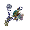
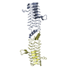
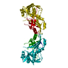
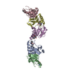
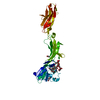
- PDB-4pkd: U1-70k in complex with U1 snRNA stem-loops 1 and U1-A RRM in comp... -
+ Open data

- Basic information
| Entry | Database: PDB / ID: 4pkd | ||||||
|---|---|---|---|---|---|---|---|
| Title | U1-70k in complex with U1 snRNA stem-loops 1 and U1-A RRM in complex with stem-loop 2 | ||||||
 Components Components |
| ||||||
 Keywords Keywords | GENE REGULATION / U1-70k / U1 snRNP / Pre-mRNA splicing / Spliceosome | ||||||
| Function / homology |  Function and homology information Function and homology informationnegative regulation of protein refolding / mRNA Splicing - Major Pathway / U1 snRNP binding / positive regulation of mRNA splicing, via spliceosome / U1 snRNP / U2-type prespliceosome / negative regulation of chaperone-mediated autophagy / regulation of RNA splicing / cellular response to transforming growth factor beta stimulus / U1 snRNA binding ...negative regulation of protein refolding / mRNA Splicing - Major Pathway / U1 snRNP binding / positive regulation of mRNA splicing, via spliceosome / U1 snRNP / U2-type prespliceosome / negative regulation of chaperone-mediated autophagy / regulation of RNA splicing / cellular response to transforming growth factor beta stimulus / U1 snRNA binding / cellular response to retinoic acid / mRNA Splicing - Major Pathway / lysosomal lumen / spliceosomal complex / mRNA splicing, via spliceosome / cellular response to tumor necrosis factor / nuclear speck / mRNA binding / RNA binding / nucleoplasm / nucleus Similarity search - Function | ||||||
| Biological species |   Homo sapiens (human) Homo sapiens (human) | ||||||
| Method |  X-RAY DIFFRACTION / SYNCHROTRON / MOLECULAR REPLACEMENT / Resolution: 2.5 Å X-RAY DIFFRACTION / SYNCHROTRON / MOLECULAR REPLACEMENT / Resolution: 2.5 Å | ||||||
 Authors Authors | Oubridge, C. / Kondo, Y. / van Roon, A.M. / Nagai, K. | ||||||
| Funding support |  Japan, 1items Japan, 1items
| ||||||
 Citation Citation | Journal: Elife / Year: 2015 Title: Crystal structure of human U1 snRNP, a small nuclear ribonucleoprotein particle, reveals the mechanism of 5' splice site recognition. Authors: Kondo, Y. / Oubridge, C. / van Roon, A.M. / Nagai, K. #1: Journal: Nature / Year: 2009Title: Crystal structure of human spliceosomal U1 snRNP at 5.5 A resolution. Authors: Pomeranz Krummel, D.A. / Oubridge, C. / Leung, A.K. / Li, J. / Nagai, K. #2: Journal: Nature / Year: 1994Title: Crystal structure at 1.92 A resolution of the RNA-binding domain of the U1A spliceosomal protein complexed with an RNA hairpin. Authors: Oubridge, C. / Ito, N. / Evans, P.R. / Teo, C.H. / Nagai, K. #3: Journal: EMBO J. / Year: 2010Title: Functional organization of the Sm core in the crystal structure of human U1 snRNP. Authors: Weber, G. / Trowitzsch, S. / Kastner, B. / Luhrmann, R. / Wahl, M.C. | ||||||
| History |
|
- Structure visualization
| Structure viewer | Molecule: MolmilJmol/JSmol |
|---|
- Downloads & links
-Download
| PDBx/mmCIF format | 4pkd.cif.gz | 103.2 KB | Display | PDBx/mmCIF format |
|---|---|---|---|---|
| PDB format | pdb4pkd.ent.gz | 73.1 KB | Display | PDB format |
| PDBx/mmJSON format | 4pkd.json.gz | Tree view | PDBx/mmJSON format | |
| Others |  Other downloads Other downloads |
-Validation report
| Arichive directory | https://data.pdbj.org/pub/pdb/validation_reports/pk/4pkdftp://data.pdbj.org/pub/pdb/validation_reports/pk/4pkd | HTTPS FTP |
|---|
-Related structure data
| Related structure data | 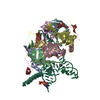 4pjoC 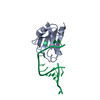 1urnS S: Starting model for refinement C: citing same article ( |
|---|---|
| Similar structure data |


-Links
 PDBj
PDBj


- Assembly
| Deposited unit | 
| ||||||||
|---|---|---|---|---|---|---|---|---|---|
| 1 |
| ||||||||
| Unit cell |
|
-Components
| #1: RNA chain | Mass: 17786.424 Da / Num. of mol.: 1 / Source method: obtained synthetically / Details: See compound details. / Source: (synth.) Homo sapiens (human) | ||||
|---|---|---|---|---|---|
| #2: Protein | Mass: 32183.576 Da / Num. of mol.: 1 / Mutation: A2G Source method: isolated from a genetically manipulated source Details: Both this protein and U1-70k are expressed together as a fusion protein. The C-terminus of U1-A and the linker peptide are disordered in the structure. The two proteins have been given ...Details: Both this protein and U1-70k are expressed together as a fusion protein. The C-terminus of U1-A and the linker peptide are disordered in the structure. The two proteins have been given separate chain identifiers so that their normal numbering is preserved., U1-A and this protein are expressed together as a fusion protein. The C-terminus of U1-A and the linker peptide are disordered in the structure. The two proteins have been given separate chain identifiers so that their normal numbering is preserved. Source: (gene. exp.) Homo sapiens (human)Gene: SNRPA, SNRNP70, RNPU1Z, RPU1, SNRP70, U1AP1 / Plasmid: pET13 Details (production host): Expresses U1-A/U1-70k fusion protein Production host:  | ||||
| #3: Chemical |   Mass: 24.305 Da / Num. of mol.: 3 / Source method: obtained synthetically / Formula: Mg Mass: 24.305 Da / Num. of mol.: 3 / Source method: obtained synthetically / Formula: Mg#4: Chemical | ChemComp-IMD / |   Mass: 69.085 Da / Num. of mol.: 1 / Source method: obtained synthetically / Formula: C3H5N2 Mass: 69.085 Da / Num. of mol.: 1 / Source method: obtained synthetically / Formula: C3H5N2#5: Water | ChemComp-HOH / |  Mass: 18.015 Da / Num. of mol.: 113 / Source method: isolated from a natural source / Formula: H2O Mass: 18.015 Da / Num. of mol.: 113 / Source method: isolated from a natural source / Formula: H2O |
-Experimental details
-Experiment
| Experiment | Method: X-RAY DIFFRACTION |
|---|
- Sample preparation
| Crystal | Density Matthews: 2.34 Å3/Da / Density % sol: 53 % / Description: Six-sided plates |
|---|---|
| Crystal grow | Temperature: 293 K / Method: vapor diffusion, sitting drop / pH: 4.2 Details: 40% MPD, 0.15 M sodium chloride, 0.1 M sodium acetate, pH 4.2 |
-Data collection
| Diffraction | Mean temperature: 100 K |
|---|---|
| Diffraction source | Source: SYNCHROTRON / Site: Diamond  / Beamline: I03 / Wavelength: 1.0332 Å / Beamline: I03 / Wavelength: 1.0332 Å |
| Detector | Type: PSI PILATUS 6M / Detector: PIXEL / Date: Oct 22, 2011 |
| Radiation | Monochromator: Double-crystal / Protocol: SINGLE WAVELENGTH / Monochromatic (M) / Laue (L): M / Scattering type: x-ray |
| Radiation wavelength | Wavelength: 1.0332 Å / Relative weight: 1 |
| Reflection | Resolution: 2.5→87.54 Å / Num. all: 16115 / Num. obs: 15967 / % possible obs: 99.1 % / Redundancy: 3.7 % / Rmerge(I) obs: 0.076 / Rsym value: 0.047 / Net I/σ(I): 11 |
| Reflection shell | Resolution: 2.5→2.64 Å / Redundancy: 3.7 % / Rmerge(I) obs: 0.35 / Mean I/σ(I) obs: 3.2 / % possible all: 99.4 |
- Processing
| Software | Name: REFMAC / Version: 5.8.0071 / Classification: refinement | ||||||||||||||||||||||||||||||||||||||||||||||||||||||||||||||||||||||||||||||||||||||||||||||||||||||||||||||||||||||||||||||||||||||||||||||||||||||||||||||||||||||||||||||||||||||
|---|---|---|---|---|---|---|---|---|---|---|---|---|---|---|---|---|---|---|---|---|---|---|---|---|---|---|---|---|---|---|---|---|---|---|---|---|---|---|---|---|---|---|---|---|---|---|---|---|---|---|---|---|---|---|---|---|---|---|---|---|---|---|---|---|---|---|---|---|---|---|---|---|---|---|---|---|---|---|---|---|---|---|---|---|---|---|---|---|---|---|---|---|---|---|---|---|---|---|---|---|---|---|---|---|---|---|---|---|---|---|---|---|---|---|---|---|---|---|---|---|---|---|---|---|---|---|---|---|---|---|---|---|---|---|---|---|---|---|---|---|---|---|---|---|---|---|---|---|---|---|---|---|---|---|---|---|---|---|---|---|---|---|---|---|---|---|---|---|---|---|---|---|---|---|---|---|---|---|---|---|---|---|---|
| Refinement | Method to determine structure: MOLECULAR REPLACEMENT Starting model: 1URN Resolution: 2.5→87 Å / Cor.coef. Fo:Fc: 0.947 / Cor.coef. Fo:Fc free: 0.893 / SU B: 10.404 / SU ML: 0.232 / Cross valid method: THROUGHOUT / ESU R: 0.618 / ESU R Free: 0.305 / Stereochemistry target values: MAXIMUM LIKELIHOOD / Details: HYDROGENS HAVE BEEN ADDED IN THE RIDING POSITIONS
| ||||||||||||||||||||||||||||||||||||||||||||||||||||||||||||||||||||||||||||||||||||||||||||||||||||||||||||||||||||||||||||||||||||||||||||||||||||||||||||||||||||||||||||||||||||||
| Solvent computation | Ion probe radii: 0.8 Å / Shrinkage radii: 0.8 Å / VDW probe radii: 1.2 Å / Solvent model: MASK | ||||||||||||||||||||||||||||||||||||||||||||||||||||||||||||||||||||||||||||||||||||||||||||||||||||||||||||||||||||||||||||||||||||||||||||||||||||||||||||||||||||||||||||||||||||||
| Displacement parameters | Biso mean: 50.252 Å2
| ||||||||||||||||||||||||||||||||||||||||||||||||||||||||||||||||||||||||||||||||||||||||||||||||||||||||||||||||||||||||||||||||||||||||||||||||||||||||||||||||||||||||||||||||||||||
| Refinement step | Cycle: 1 / Resolution: 2.5→87 Å
| ||||||||||||||||||||||||||||||||||||||||||||||||||||||||||||||||||||||||||||||||||||||||||||||||||||||||||||||||||||||||||||||||||||||||||||||||||||||||||||||||||||||||||||||||||||||
| Refine LS restraints |
|