 Movie
Movie Controller

[English] 日本語
 Yorodumi
- PDB-4pbx: Crystal structure of the six N-terminal domains of human receptor... -
+ Open data

- Basic information
| Entry | Database: PDB / ID: 4pbx | ||||||||||||||||||
|---|---|---|---|---|---|---|---|---|---|---|---|---|---|---|---|---|---|---|---|
| Title | Crystal structure of the six N-terminal domains of human receptor protein tyrosine phosphatase sigma | ||||||||||||||||||
 Components Components | Receptor-type tyrosine-protein phosphatase S | ||||||||||||||||||
 Keywords Keywords | HYDROLASE / Signaling protein / Synapse Cell signalling Cell surface receptor | ||||||||||||||||||
| Function / homology |  Function and homology information Function and homology informationnegative regulation of toll-like receptor 9 signaling pathway / Signaling by NTRK3 (TRKC) / trans-synaptic signaling / negative regulation of interferon-alpha production / chondroitin sulfate binding / Receptor-type tyrosine-protein phosphatases / establishment of endothelial intestinal barrier / negative regulation of collateral sprouting / negative regulation of dendritic spine development / synaptic membrane adhesion ...negative regulation of toll-like receptor 9 signaling pathway / Signaling by NTRK3 (TRKC) / trans-synaptic signaling / negative regulation of interferon-alpha production / chondroitin sulfate binding / Receptor-type tyrosine-protein phosphatases / establishment of endothelial intestinal barrier / negative regulation of collateral sprouting / negative regulation of dendritic spine development / synaptic membrane adhesion / corpus callosum development / negative regulation of axon extension / regulation of postsynaptic density assembly / negative regulation of axon regeneration / Synaptic adhesion-like molecules / negative regulation of interferon-beta production / heparan sulfate proteoglycan binding / protein dephosphorylation / spinal cord development / peptidyl-tyrosine dephosphorylation / ECM proteoglycans / phosphoprotein phosphatase activity / protein-tyrosine-phosphatase / protein tyrosine phosphatase activity / cerebellum development / hippocampus development / cerebral cortex development / postsynaptic density membrane / modulation of chemical synaptic transmission / Schaffer collateral - CA1 synapse / negative regulation of neuron projection development / heparin binding / synaptic vesicle membrane / growth cone / presynaptic membrane / perikaryon / axon / glutamatergic synapse / signal transduction / extracellular exosome / plasma membrane / cytosol Similarity search - Function | ||||||||||||||||||
| Biological species |  Homo sapiens (human) Homo sapiens (human) | ||||||||||||||||||
| Method |  X-RAY DIFFRACTION / SYNCHROTRON / MOLECULAR REPLACEMENT / Resolution: 3.15 Å X-RAY DIFFRACTION / SYNCHROTRON / MOLECULAR REPLACEMENT / Resolution: 3.15 Å | ||||||||||||||||||
 Authors Authors | Coles, C.H. / Mitakidis, N. / Zhang, P. / Elegheert, J. / Lu, W. / Stoker, A.W. / Nakagawa, T. / Craig, A.M. / Jones, E.Y. / Aricescu, A.R. | ||||||||||||||||||
| Funding support |  United Kingdom, 5items United Kingdom, 5items
| ||||||||||||||||||
 Citation Citation | Journal: Nat Commun / Year: 2014 Title: Structural basis for extracellular cis and trans RPTP sigma signal competition in synaptogenesis. Authors: Coles, C.H. / Mitakidis, N. / Zhang, P. / Elegheert, J. / Lu, W. / Stoker, A.W. / Nakagawa, T. / Craig, A.M. / Jones, E.Y. / Aricescu, A.R. | ||||||||||||||||||
| History |
|
- Structure visualization
| Structure viewer | Molecule: MolmilJmol/JSmol |
|---|
- Downloads & links
-Download
| PDBx/mmCIF format | 4pbx.cif.gz | 239.3 KB | Display | PDBx/mmCIF format |
|---|---|---|---|---|
| PDB format | pdb4pbx.ent.gz | 194 KB | Display | PDB format |
| PDBx/mmJSON format | 4pbx.json.gz | Tree view | PDBx/mmJSON format | |
| Others |  Other downloads Other downloads |
-Validation report
| Arichive directory | https://data.pdbj.org/pub/pdb/validation_reports/pb/4pbxftp://data.pdbj.org/pub/pdb/validation_reports/pb/4pbx | HTTPS FTP |
|---|
-Related structure data
| Related structure data | 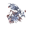 4pbvC 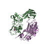 4pbwC 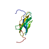 2djuS  2yd3S  2yd4S 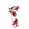 2yd9S S: Starting model for refinement C: citing same article ( |
|---|---|
| Similar structure data |


-Links
 PDBj
PDBj


- Assembly
| Deposited unit | 
| ||||||||
|---|---|---|---|---|---|---|---|---|---|
| 1 |
| ||||||||
| Unit cell |
|
-Components
| #1: Protein | Mass: 64344.035 Da / Num. of mol.: 1 / Fragment: Residues 30-588 Source method: isolated from a genetically manipulated source Source: (gene. exp.) Homo sapiens (human) / Gene: PTPRS / Plasmid: pHLsec / Cell line (production host): HEK293T / Production host: Homo sapiens (human) / References: UniProt: Q13332, protein-tyrosine-phosphatase | ||||
|---|---|---|---|---|---|
| #2: Sugar |   Type: D-saccharide, beta linking / Mass: 221.208 Da / Num. of mol.: 2 / Source method: obtained synthetically / Formula: C8H15NO6 Type: D-saccharide, beta linking / Mass: 221.208 Da / Num. of mol.: 2 / Source method: obtained synthetically / Formula: C8H15NO6Compound details | The sample sequence matches to isoform 6 of UniProt Q13332 | Has protein modification | Y | |
-Experimental details
-Experiment
| Experiment | Method: X-RAY DIFFRACTION |
|---|
- Sample preparation
| Crystal | Density Matthews: 5.87 Å3/Da / Density % sol: 79.04 % |
|---|---|
| Crystal grow | Temperature: 293.5 K / Method: vapor diffusion, sitting drop / pH: 6 Details: 10 % PEG 400, 0.01 M magnesium chloride, 0.1 M potassium chloride, 0.05 M MES pH 6.0 |
-Data collection
| Diffraction | Mean temperature: 100 K |
|---|---|
| Diffraction source | Source: SYNCHROTRON / Site: ESRF  / Beamline: ID29 / Wavelength: 0.9788 Å / Beamline: ID29 / Wavelength: 0.9788 Å |
| Detector | Type: ADSC QUANTUM 315r / Detector: CCD / Date: Sep 8, 2008 |
| Radiation | Protocol: SINGLE WAVELENGTH / Monochromatic (M) / Laue (L): M / Scattering type: x-ray |
| Radiation wavelength | Wavelength: 0.9788 Å / Relative weight: 1 |
| Reflection | Resolution: 3.15→99.4 Å / Num. obs: 25619 / % possible obs: 95.6 % / Redundancy: 7.6 % / Rmerge(I) obs: 0.078 / Net I/σ(I): 17.8 |
| Reflection shell | Resolution: 3.15→3.23 Å / Redundancy: 7.5 % / Rmerge(I) obs: 0.991 / Mean I/σ(I) obs: 2.1 / % possible all: 96.1 |
- Processing
| Software |
| ||||||||||||||||||||||||||||||||||||||||||||||||||||||||||||||||||||||||||||||||||||||||||||||||||||||||||||||||||||||||||||||||||||||||||||||||||||||||||||||||||||||||||||||||||||||
|---|---|---|---|---|---|---|---|---|---|---|---|---|---|---|---|---|---|---|---|---|---|---|---|---|---|---|---|---|---|---|---|---|---|---|---|---|---|---|---|---|---|---|---|---|---|---|---|---|---|---|---|---|---|---|---|---|---|---|---|---|---|---|---|---|---|---|---|---|---|---|---|---|---|---|---|---|---|---|---|---|---|---|---|---|---|---|---|---|---|---|---|---|---|---|---|---|---|---|---|---|---|---|---|---|---|---|---|---|---|---|---|---|---|---|---|---|---|---|---|---|---|---|---|---|---|---|---|---|---|---|---|---|---|---|---|---|---|---|---|---|---|---|---|---|---|---|---|---|---|---|---|---|---|---|---|---|---|---|---|---|---|---|---|---|---|---|---|---|---|---|---|---|---|---|---|---|---|---|---|---|---|---|---|
| Refinement | Method to determine structure: MOLECULAR REPLACEMENT Starting model: 2YD3, 2YD4, 2YD9, 2DJU Resolution: 3.15→172.15 Å / Cor.coef. Fo:Fc: 0.91 / Cor.coef. Fo:Fc free: 0.9 / SU B: 42.451 / SU ML: 0.312 / Cross valid method: THROUGHOUT / ESU R: 0.622 / ESU R Free: 0.369 / Stereochemistry target values: MAXIMUM LIKELIHOOD / Details: HYDROGENS HAVE BEEN ADDED IN THE RIDING POSITIONS
| ||||||||||||||||||||||||||||||||||||||||||||||||||||||||||||||||||||||||||||||||||||||||||||||||||||||||||||||||||||||||||||||||||||||||||||||||||||||||||||||||||||||||||||||||||||||
| Solvent computation | Ion probe radii: 0.8 Å / Shrinkage radii: 0.8 Å / VDW probe radii: 1.2 Å / Solvent model: MASK | ||||||||||||||||||||||||||||||||||||||||||||||||||||||||||||||||||||||||||||||||||||||||||||||||||||||||||||||||||||||||||||||||||||||||||||||||||||||||||||||||||||||||||||||||||||||
| Displacement parameters | Biso mean: 120.828 Å2
| ||||||||||||||||||||||||||||||||||||||||||||||||||||||||||||||||||||||||||||||||||||||||||||||||||||||||||||||||||||||||||||||||||||||||||||||||||||||||||||||||||||||||||||||||||||||
| Refinement step | Cycle: 1 / Resolution: 3.15→172.15 Å
| ||||||||||||||||||||||||||||||||||||||||||||||||||||||||||||||||||||||||||||||||||||||||||||||||||||||||||||||||||||||||||||||||||||||||||||||||||||||||||||||||||||||||||||||||||||||
| Refine LS restraints |
|