 Movie
Movie Controller

[English] 日本語
 Yorodumi
- PDB-4efa: Crystal Structure of the Heterotrimeric EGChead Peripheral Stalk ... -
+ Open data

- Basic information
| Entry | Database: PDB / ID: 4efa | ||||||
|---|---|---|---|---|---|---|---|
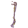
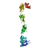
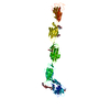
| Title | Crystal Structure of the Heterotrimeric EGChead Peripheral Stalk Complex of the Yeast Vacuolar ATPase - second conformation | ||||||
 Components Components |
| ||||||
 Keywords Keywords | HYDROLASE / heterotrimer / peripheral stalk / vacuolar ATPase | ||||||
| Function / homology |  Function and homology information Function and homology informationInsulin receptor recycling / Transferrin endocytosis and recycling / ROS and RNS production in phagocytes / Golgi lumen acidification / Amino acids regulate mTORC1 / vacuolar proton-transporting V-type ATPase, V1 domain / endosomal lumen acidification / vacuolar acidification / fungal-type vacuole membrane / proton-transporting ATPase activity, rotational mechanism ...Insulin receptor recycling / Transferrin endocytosis and recycling / ROS and RNS production in phagocytes / Golgi lumen acidification / Amino acids regulate mTORC1 / vacuolar proton-transporting V-type ATPase, V1 domain / endosomal lumen acidification / vacuolar acidification / fungal-type vacuole membrane / proton-transporting ATPase activity, rotational mechanism / proton transmembrane transport / membrane raft / Golgi membrane / ATP binding Similarity search - Function | ||||||
| Biological species |  | ||||||
| Method |  X-RAY DIFFRACTION / SYNCHROTRON / MOLECULAR REPLACEMENT / molecular replacement / Resolution: 2.8163 Å X-RAY DIFFRACTION / SYNCHROTRON / MOLECULAR REPLACEMENT / molecular replacement / Resolution: 2.8163 Å | ||||||
 Authors Authors | Oot, R.A. / Huang, L.S. / Berry, E.A. / Wilkens, S. | ||||||
 Citation Citation | Journal: Structure / Year: 2012 Title: Crystal Structure of the Yeast Vacuolar ATPase Heterotrimeric EGC(head) Peripheral Stalk Complex. Authors: Oot, R.A. / Huang, L.S. / Berry, E.A. / Wilkens, S. | ||||||
| History |
|
- Structure visualization
| Structure viewer | Molecule: MolmilJmol/JSmol |
|---|
- Downloads & links
-Download
| PDBx/mmCIF format | 4efa.cif.gz | 189 KB | Display | PDBx/mmCIF format |
|---|---|---|---|---|
| PDB format | pdb4efa.ent.gz | 151.8 KB | Display | PDB format |
| PDBx/mmJSON format | 4efa.json.gz | Tree view | PDBx/mmJSON format | |
| Others |  Other downloads Other downloads |
-Validation report
| Arichive directory | https://data.pdbj.org/pub/pdb/validation_reports/ef/4efaftp://data.pdbj.org/pub/pdb/validation_reports/ef/4efa | HTTPS FTP |
|---|
-Related structure data
| Related structure data |  4dl0SC S: Starting model for refinement C: citing same article ( |
|---|---|
| Similar structure data |


-Links
 PDBj
PDBj

- Assembly
| Deposited unit | 
| ||||||||
|---|---|---|---|---|---|---|---|---|---|
| 1 |
| ||||||||
| Unit cell |
|
-Components
| #1: Protein | Mass: 14586.475 Da / Num. of mol.: 1 / Fragment: UNP Residues 158-277 Source method: isolated from a genetically manipulated source Source: (gene. exp.) Strain: ATCC 204508 / S288c / Gene: VAT3, VATC, VMA5, YKL080W, YKL410 / Plasmid: pMALc2e / Production host:  References: UniProt: P31412, H+-transporting two-sector ATPase | ||
|---|---|---|---|
| #2: Protein | Mass: 13218.298 Da / Num. of mol.: 1 Source method: isolated from a genetically manipulated source Source: (gene. exp.) Strain: ATCC 204508 / S288c / Gene: VMA10, YHR039BC, YHR039C-A, YHR039C-B / Plasmid: pMALc2e / Production host: References: UniProt: P48836, H+-transporting two-sector ATPase | ||
| #3: Protein | Mass: 26508.393 Da / Num. of mol.: 1 Source method: isolated from a genetically manipulated source Source: (gene. exp.) Strain: ATCC 204508 / S288c / Gene: O6241, VAT5, VMA4, YOR332W / Plasmid: pMALc2e / Production host: References: UniProt: P22203, H+-transporting two-sector ATPase | ||
| #4: Chemical | ChemComp-SO4 /   Mass: 96.063 Da / Num. of mol.: 4 / Source method: obtained synthetically / Formula: SO4 Mass: 96.063 Da / Num. of mol.: 4 / Source method: obtained synthetically / Formula: SO4#5: Water | ChemComp-HOH / |  Mass: 18.015 Da / Num. of mol.: 24 / Source method: isolated from a natural source / Formula: H2O Mass: 18.015 Da / Num. of mol.: 24 / Source method: isolated from a natural source / Formula: H2O |
-Experimental details
-Experiment
| Experiment | Method: X-RAY DIFFRACTION / Number of used crystals: 1 |
|---|
- Sample preparation
| Crystal | Density Matthews: 2.6 Å3/Da / Density % sol: 52.63 % |
|---|---|
| Crystal grow | Temperature: 292 K / Method: vapor diffusion, hanging drop / pH: 6 Details: 0.1 M lithium sulfate, 0.1 M MES, 20% PEG mme 2000, 0.15 M glycine , pH 6, VAPOR DIFFUSION, HANGING DROP, temperature 292K |
-Data collection
| Diffraction | Mean temperature: 100 K |
|---|---|
| Diffraction source | Source: SYNCHROTRON / Site: CHESS  / Beamline: F1 / Wavelength: 0.918 Å / Beamline: F1 / Wavelength: 0.918 Å |
| Detector | Type: ADSC QUANTUM 270 / Detector: CCD / Date: Oct 22, 2011 |
| Diffraction measurement | Details: 1.00 degrees, 15.0 sec, detector distance 380.00 mm Method: omega scans |
| Radiation | Monochromator: Si(111) / Protocol: SINGLE WAVELENGTH / Monochromatic (M) / Laue (L): M / Scattering type: x-ray |
| Radiation wavelength | Wavelength: 0.918 Å / Relative weight: 1 |
| Reflection | Av R equivalents: 0.143 / Number: 198487 |
| Reflection | Resolution: 2.8→40 Å / Num. all: 14238 / Num. obs: 14187 / % possible obs: 99.9 % / Observed criterion σ(F): 0 / Observed criterion σ(I): -3 / Redundancy: 13.9 % / Rmerge(I) obs: 0.132 / Rsym value: 0.132 / Net I/σ(I): 35.089 |
| Reflection shell | Resolution: 2.8→2.85 Å / Redundancy: 14.1 % / Rmerge(I) obs: 0 / Mean I/σ(I) obs: 2.306 / Rsym value: 0 / % possible all: 100 |
| Cell measurement | Reflection used: 198487 |
-Phasing
| Phasing | Method: molecular replacement | |||||||||
|---|---|---|---|---|---|---|---|---|---|---|
| Phasing MR |
|
- Processing
| Software |
| ||||||||||||||||||||||||||||||||||||||||||||||||||||||||||||||||||||||||||||||||||||||||||||||||||||||||||||||||||||||||||||||||||||||||||||||||||||||||||||||||||||||||||||||||||||||||||||||||||||||||||||||||||||||||||||||||||||||||||||||||||||||||||
|---|---|---|---|---|---|---|---|---|---|---|---|---|---|---|---|---|---|---|---|---|---|---|---|---|---|---|---|---|---|---|---|---|---|---|---|---|---|---|---|---|---|---|---|---|---|---|---|---|---|---|---|---|---|---|---|---|---|---|---|---|---|---|---|---|---|---|---|---|---|---|---|---|---|---|---|---|---|---|---|---|---|---|---|---|---|---|---|---|---|---|---|---|---|---|---|---|---|---|---|---|---|---|---|---|---|---|---|---|---|---|---|---|---|---|---|---|---|---|---|---|---|---|---|---|---|---|---|---|---|---|---|---|---|---|---|---|---|---|---|---|---|---|---|---|---|---|---|---|---|---|---|---|---|---|---|---|---|---|---|---|---|---|---|---|---|---|---|---|---|---|---|---|---|---|---|---|---|---|---|---|---|---|---|---|---|---|---|---|---|---|---|---|---|---|---|---|---|---|---|---|---|---|---|---|---|---|---|---|---|---|---|---|---|---|---|---|---|---|---|---|---|---|---|---|---|---|---|---|---|---|---|---|---|---|---|---|---|---|---|---|---|---|---|---|---|---|---|---|---|---|---|
| Refinement | Method to determine structure: MOLECULAR REPLACEMENT Starting model: PDB ENTRY 4DL0 Resolution: 2.8163→38.655 Å / Occupancy max: 1 / Occupancy min: 1 / SU ML: 0.38 / σ(F): 1.34 / Phase error: 30.19 / Stereochemistry target values: ML
| ||||||||||||||||||||||||||||||||||||||||||||||||||||||||||||||||||||||||||||||||||||||||||||||||||||||||||||||||||||||||||||||||||||||||||||||||||||||||||||||||||||||||||||||||||||||||||||||||||||||||||||||||||||||||||||||||||||||||||||||||||||||||||
| Solvent computation | Shrinkage radii: 0.73 Å / VDW probe radii: 1 Å / Solvent model: FLAT BULK SOLVENT MODEL / Bsol: 58.287 Å2 / ksol: 0.327 e/Å3 | ||||||||||||||||||||||||||||||||||||||||||||||||||||||||||||||||||||||||||||||||||||||||||||||||||||||||||||||||||||||||||||||||||||||||||||||||||||||||||||||||||||||||||||||||||||||||||||||||||||||||||||||||||||||||||||||||||||||||||||||||||||||||||
| Displacement parameters | Biso max: 222.28 Å2 / Biso mean: 93.5261 Å2 / Biso min: 22.53 Å2
| ||||||||||||||||||||||||||||||||||||||||||||||||||||||||||||||||||||||||||||||||||||||||||||||||||||||||||||||||||||||||||||||||||||||||||||||||||||||||||||||||||||||||||||||||||||||||||||||||||||||||||||||||||||||||||||||||||||||||||||||||||||||||||
| Refinement step | Cycle: LAST / Resolution: 2.8163→38.655 Å
| ||||||||||||||||||||||||||||||||||||||||||||||||||||||||||||||||||||||||||||||||||||||||||||||||||||||||||||||||||||||||||||||||||||||||||||||||||||||||||||||||||||||||||||||||||||||||||||||||||||||||||||||||||||||||||||||||||||||||||||||||||||||||||
| Refine LS restraints |
| ||||||||||||||||||||||||||||||||||||||||||||||||||||||||||||||||||||||||||||||||||||||||||||||||||||||||||||||||||||||||||||||||||||||||||||||||||||||||||||||||||||||||||||||||||||||||||||||||||||||||||||||||||||||||||||||||||||||||||||||||||||||||||
| LS refinement shell | Refine-ID: X-RAY DIFFRACTION / Total num. of bins used: 10
| ||||||||||||||||||||||||||||||||||||||||||||||||||||||||||||||||||||||||||||||||||||||||||||||||||||||||||||||||||||||||||||||||||||||||||||||||||||||||||||||||||||||||||||||||||||||||||||||||||||||||||||||||||||||||||||||||||||||||||||||||||||||||||
| Refinement TLS params. | Method: refined / Refine-ID: X-RAY DIFFRACTION
| ||||||||||||||||||||||||||||||||||||||||||||||||||||||||||||||||||||||||||||||||||||||||||||||||||||||||||||||||||||||||||||||||||||||||||||||||||||||||||||||||||||||||||||||||||||||||||||||||||||||||||||||||||||||||||||||||||||||||||||||||||||||||||
| Refinement TLS group |
|
