 Movie
Movie Controller

[English] 日本語
 Yorodumi
- PDB-6h3a: Crystal structure of the KAP1 RBCC domain in complex with the SMA... -
+ Open data

- Basic information
| Entry | Database: PDB / ID: 6h3a | ||||||
|---|---|---|---|---|---|---|---|
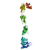
| Title | Crystal structure of the KAP1 RBCC domain in complex with the SMARCAD1 CUE1 domain. | ||||||
 Components Components |
| ||||||
 Keywords Keywords | LIGASE / TRIM28 / transcriptional co-repressor / CUE domain / Ubuiquitin | ||||||
| Function / homology |  Function and homology information Function and homology informationATP-dependent histone chaperone activity / convergent extension involved in axis elongation / Krueppel-associated box domain binding / embryonic placenta morphogenesis / negative regulation of single stranded viral RNA replication via double stranded DNA intermediate / regulation of DNA recombination / chromosome separation / chromo shadow domain binding / suppression of viral release by host / DNA double-strand break processing ...ATP-dependent histone chaperone activity / convergent extension involved in axis elongation / Krueppel-associated box domain binding / embryonic placenta morphogenesis / negative regulation of single stranded viral RNA replication via double stranded DNA intermediate / regulation of DNA recombination / chromosome separation / chromo shadow domain binding / suppression of viral release by host / DNA double-strand break processing / genomic imprinting / Generic Transcription Pathway / nucleosome array spacer activity / SUMO transferase activity / ATP-dependent chromatin remodeler activity / DNA methylation-dependent constitutive heterochromatin formation / nuclear replication fork / epithelial to mesenchymal transition / protein sumoylation / heterochromatin / embryo implantation / SUMOylation of transcription cofactors / positive regulation of DNA repair / DNA helicase activity / Regulation of endogenous retroelements by KRAB-ZFP proteins / ubiquitin binding / promoter-specific chromatin binding / euchromatin / positive regulation of protein import into nucleus / RING-type E3 ubiquitin transferase / Hydrolases; Acting on acid anhydrides; Acting on acid anhydrides to facilitate cellular and subcellular movement / RNA polymerase II transcription regulator complex / HCMV Early Events / ubiquitin-protein transferase activity / transcription corepressor activity / ubiquitin protein ligase activity / heterochromatin formation / site of double-strand break / chromatin organization / proteasome-mediated ubiquitin-dependent protein catabolic process / protein kinase activity / transcription coactivator activity / chromatin remodeling / innate immune response / DNA repair / negative regulation of DNA-templated transcription / chromatin binding / ubiquitin protein ligase binding / positive regulation of DNA-templated transcription / chromatin / negative regulation of transcription by RNA polymerase II / ATP hydrolysis activity / positive regulation of transcription by RNA polymerase II / protein-containing complex / DNA binding / RNA binding / zinc ion binding / nucleoplasm / ATP binding / nucleus Similarity search - Function | ||||||
| Biological species |  Homo sapiens (human) Homo sapiens (human) | ||||||
| Method |  X-RAY DIFFRACTION / SYNCHROTRON / SAD / Resolution: 5.505 Å X-RAY DIFFRACTION / SYNCHROTRON / SAD / Resolution: 5.505 Å | ||||||
 Authors Authors | Newman, J.A. / Aitkenhead, H. / Lim, M. / Williams, H.L. / Svejstrup, J.Q. / von Delft, F. / Arrowsmith, C.H. / Edwards, A. / Bountra, C. / Gileadi, O. | ||||||
 Citation Citation | Journal: Structure / Year: 2019 Title: A Ubiquitin-Binding Domain that Binds a Structural Fold Distinct from that of Ubiquitin. Authors: Lim, M. / Newman, J.A. / Williams, H.L. / Masino, L. / Aitkenhead, H. / Gravard, A.E. / Gileadi, O. / Svejstrup, J.Q. | ||||||
| History |
|
- Structure visualization
| Structure viewer | Molecule: MolmilJmol/JSmol |
|---|
- Downloads & links
-Download
| PDBx/mmCIF format | 6h3a.cif.gz | 150.5 KB | Display | PDBx/mmCIF format |
|---|---|---|---|---|
| PDB format | pdb6h3a.ent.gz | 112.1 KB | Display | PDB format |
| PDBx/mmJSON format | 6h3a.json.gz | Tree view | PDBx/mmJSON format | |
| Others |  Other downloads Other downloads |
-Validation report
| Arichive directory | https://data.pdbj.org/pub/pdb/validation_reports/h3/6h3aftp://data.pdbj.org/pub/pdb/validation_reports/h3/6h3a | HTTPS FTP |
|---|
-Related structure data


-Links
 PDBj
PDBj

- Assembly
| Deposited unit | 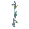
| ||||||||
|---|---|---|---|---|---|---|---|---|---|
| 1 |
| ||||||||
| Unit cell |
|
-Components
| #1: Protein | Mass: 28799.678 Da / Num. of mol.: 2 Source method: isolated from a genetically manipulated source Source: (gene. exp.) Homo sapiens (human) / Gene: SMARCAD1, KIAA1122 / Production host:  #2: Protein | Mass: 42501.395 Da / Num. of mol.: 2 Source method: isolated from a genetically manipulated source Source: (gene. exp.) Homo sapiens (human) / Gene: TRIM28, KAP1, RNF96, TIF1B / Production host: References: UniProt: Q13263, RING-type E3 ubiquitin transferase #3: Chemical | ChemComp-ZN /   Mass: 65.409 Da / Num. of mol.: 8 / Source method: obtained synthetically / Formula: Zn Mass: 65.409 Da / Num. of mol.: 8 / Source method: obtained synthetically / Formula: Zn |
|---|
-Experimental details
-Experiment
| Experiment | Method: X-RAY DIFFRACTION / Number of used crystals: 1 |
|---|
- Sample preparation
| Crystal grow | Temperature: 277 K / Method: vapor diffusion, sitting drop Details: 1.2 M Sodium Malonate, 0.5 % Jeffamine ED-2003 and 0.1 M HEPES pH 7.0 |
|---|
-Data collection
| Diffraction | Mean temperature: 100 K / Serial crystal experiment: N |
|---|---|
| Diffraction source | Source: SYNCHROTRON / Site: Diamond  / Beamline: I03 / Wavelength: 0.976 Å / Beamline: I03 / Wavelength: 0.976 Å |
| Detector | Type: DECTRIS PILATUS3 S 6M / Detector: PIXEL / Date: Sep 21, 2017 |
| Radiation | Protocol: SINGLE WAVELENGTH / Monochromatic (M) / Laue (L): M / Scattering type: x-ray |
| Radiation wavelength | Wavelength: 0.976 Å / Relative weight: 1 |
| Reflection | Resolution: 5.5→80.1 Å / Num. obs: 14734 / % possible obs: 100 % / Redundancy: 19.7 % / Biso Wilson estimate: 280 Å2 / CC1/2: 0.999 / Rmerge(I) obs: 0.155 / Rpim(I) all: 0.04 / Net I/σ(I): 11.5 |
| Reflection shell | Resolution: 5.5→6.15 Å / Redundancy: 19.5 % / Rmerge(I) obs: 1.25 / Mean I/σ(I) obs: 2.6 / Num. unique obs: 3240 / CC1/2: 0.16 / Rpim(I) all: 0.29 / % possible all: 100 |
- Processing
| Software |
| ||||||||||||||||||||||||
|---|---|---|---|---|---|---|---|---|---|---|---|---|---|---|---|---|---|---|---|---|---|---|---|---|---|
| Refinement | Method to determine structure: SAD / Resolution: 5.505→74.978 Å / SU ML: 1.53 / Cross valid method: FREE R-VALUE / σ(F): 1.49 / Phase error: 45.15
| ||||||||||||||||||||||||
| Solvent computation | Shrinkage radii: 0.9 Å / VDW probe radii: 1.11 Å | ||||||||||||||||||||||||
| Refinement step | Cycle: LAST / Resolution: 5.505→74.978 Å
| ||||||||||||||||||||||||
| Refine LS restraints |
| ||||||||||||||||||||||||
| LS refinement shell | Highest resolution: 5.505 Å |
