 Movie
Movie Controller

[English] 日本語
 Yorodumi
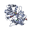
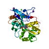
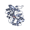
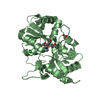
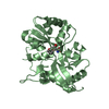
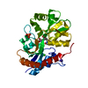
- PDB-4nwd: Crystal structure of the kainate receptor GluK3 ligand-binding do... -
+ Open data

- Basic information
| Entry | Database: PDB / ID: 4nwd | ||||||
|---|---|---|---|---|---|---|---|
| Title | Crystal structure of the kainate receptor GluK3 ligand-binding domain in complex with the agonist (2S,4R)-4-(3-Methylamino-3-oxopropyl)glutamic acid at 2.6 A resolution | ||||||
 Components Components | Glutamate receptor ionotropic, kainate 3 | ||||||
 Keywords Keywords | MEMBRANE PROTEIN/AGONIST / kainate receptor / ligand binding domain / agonist / ionotropic glutamate receptor / MEMBRABE PROTEIN-AGONIST complex / MEMBRANE PROTEIN-AGONIST complex | ||||||
| Function / homology |  Function and homology information Function and homology informationPresynaptic function of Kainate receptors / cochlear hair cell ribbon synapse / adenylate cyclase inhibiting G protein-coupled glutamate receptor activity / G protein-coupled glutamate receptor signaling pathway / Activation of Ca-permeable Kainate Receptor / kainate selective glutamate receptor complex / glutamate receptor activity / negative regulation of synaptic transmission, glutamatergic / glutamate receptor signaling pathway / kainate selective glutamate receptor activity ...Presynaptic function of Kainate receptors / cochlear hair cell ribbon synapse / adenylate cyclase inhibiting G protein-coupled glutamate receptor activity / G protein-coupled glutamate receptor signaling pathway / Activation of Ca-permeable Kainate Receptor / kainate selective glutamate receptor complex / glutamate receptor activity / negative regulation of synaptic transmission, glutamatergic / glutamate receptor signaling pathway / kainate selective glutamate receptor activity / glutamate-gated receptor activity / glutamate-gated calcium ion channel activity / dendrite cytoplasm / ligand-gated monoatomic ion channel activity involved in regulation of presynaptic membrane potential / transmitter-gated monoatomic ion channel activity involved in regulation of postsynaptic membrane potential / synaptic transmission, glutamatergic / regulation of membrane potential / postsynaptic density membrane / modulation of chemical synaptic transmission / terminal bouton / presynaptic membrane / chemical synaptic transmission / perikaryon / postsynaptic membrane / axon / dendrite / glutamatergic synapse / plasma membrane Similarity search - Function | ||||||
| Biological species |  | ||||||
| Method |  X-RAY DIFFRACTION / SYNCHROTRON / MOLECULAR REPLACEMENT / molecular replacement / Resolution: 2.6 Å X-RAY DIFFRACTION / SYNCHROTRON / MOLECULAR REPLACEMENT / molecular replacement / Resolution: 2.6 Å | ||||||
 Authors Authors | Venskutonyte, R. / Larsen, A.P. / Frydenvang, K. / Gajhede, M. / Kastrup, J.S. | ||||||
 Citation Citation | Journal: Chemmedchem / Year: 2014 Title: Molecular Recognition of Two 2,4-syn-Functionalized (S)-Glutamate Analogues by the Kainate Receptor GluK3 Ligand Binding Domain. Authors: Venskutonyte, R. / Larsen, A.P. / Frydenvang, K. / Gajhede, M. / Sagot, E. / Assaf, Z. / Gefflaut, T. / Pickering, D.S. / Bunch, L. / Kastrup, J.S. #1: Journal: J.Struct.Biol. / Year: 2011Title: Binding site and interlobe interactions of the ionotropic glutamate receptor GluK3 ligand binding domain revealed by high resolution crystal structure in complex with (S)-glutamate. Authors: Venskutonyte, R. / Frydenvang, K. / Gajhede, M. / Bunch, L. / Pickering, D.S. / Kastrup, J.S. | ||||||
| History |
|
- Structure visualization
| Structure viewer | Molecule: MolmilJmol/JSmol |
|---|
- Downloads & links
-Download
| PDBx/mmCIF format | 4nwd.cif.gz | 118.6 KB | Display | PDBx/mmCIF format |
|---|---|---|---|---|
| PDB format | pdb4nwd.ent.gz | 92.2 KB | Display | PDB format |
| PDBx/mmJSON format | 4nwd.json.gz | Tree view | PDBx/mmJSON format | |
| Others |  Other downloads Other downloads |
-Validation report
| Arichive directory | https://data.pdbj.org/pub/pdb/validation_reports/nw/4nwdftp://data.pdbj.org/pub/pdb/validation_reports/nw/4nwd | HTTPS FTP |
|---|
-Related structure data
| Related structure data | 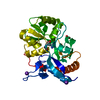 4nwcC  3s9eS S: Starting model for refinement C: citing same article ( |
|---|---|
| Similar structure data |


-Links
 PDBj
PDBj

- Assembly
| Deposited unit | 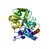
| ||||||||
|---|---|---|---|---|---|---|---|---|---|
| 1 |
| ||||||||
| Unit cell |
| ||||||||
| Components on special symmetry positions |
|
-Components
| #1: Protein | Mass: 29092.453 Da / Num. of mol.: 1 / Fragment: unp residues 432-546 and unp residues 669-806 Source method: isolated from a genetically manipulated source Source: (gene. exp.)  | ||||||
|---|---|---|---|---|---|---|---|
| #2: Chemical | ChemComp-2QD / (  Type: L-peptide linking / Mass: 232.234 Da / Num. of mol.: 1 / Source method: obtained synthetically / Formula: C9H16N2O5 Type: L-peptide linking / Mass: 232.234 Da / Num. of mol.: 1 / Source method: obtained synthetically / Formula: C9H16N2O5 | ||||||
| #3: Chemical |   Mass: 39.098 Da / Num. of mol.: 2 / Source method: obtained synthetically / Formula: K Mass: 39.098 Da / Num. of mol.: 2 / Source method: obtained synthetically / Formula: K#4: Chemical | ChemComp-CL / |   Mass: 35.453 Da / Num. of mol.: 1 / Source method: obtained synthetically / Formula: Cl Mass: 35.453 Da / Num. of mol.: 1 / Source method: obtained synthetically / Formula: Cl#5: Water | ChemComp-HOH / |  Mass: 18.015 Da / Num. of mol.: 49 / Source method: isolated from a natural source / Formula: H2O Mass: 18.015 Da / Num. of mol.: 49 / Source method: isolated from a natural source / Formula: H2OHas protein modification | Y | |
-Experimental details
-Experiment
| Experiment | Method: X-RAY DIFFRACTION / Number of used crystals: 1 |
|---|
- Sample preparation
| Crystal | Density Matthews: 2.57 Å3/Da / Density % sol: 52.11 % |
|---|---|
| Crystal grow | Temperature: 293 K / Method: vapor diffusion, hanging drop / pH: 8.2 Details: 1.8 M NaH2PO4/K2HPO4, co-crystallized with (S)-glutamate, soaked with (2S,4R)-4-(3-Methylamino-3-oxopropyl)glutamic acid, pH 8.2, VAPOR DIFFUSION, HANGING DROP, temperature 293K |
-Data collection
| Diffraction | Mean temperature: 100 K | ||||||||||||||||||||||||||||||||||||||||||||||||||||||||||||||||||||||||||||||||||||||||
|---|---|---|---|---|---|---|---|---|---|---|---|---|---|---|---|---|---|---|---|---|---|---|---|---|---|---|---|---|---|---|---|---|---|---|---|---|---|---|---|---|---|---|---|---|---|---|---|---|---|---|---|---|---|---|---|---|---|---|---|---|---|---|---|---|---|---|---|---|---|---|---|---|---|---|---|---|---|---|---|---|---|---|---|---|---|---|---|---|---|
| Diffraction source | Source: SYNCHROTRON / Site: MAX II  / Beamline: I911-3 / Wavelength: 0.9974 Å / Beamline: I911-3 / Wavelength: 0.9974 Å | ||||||||||||||||||||||||||||||||||||||||||||||||||||||||||||||||||||||||||||||||||||||||
| Detector | Type: MARMOSAIC 225 mm CCD / Detector: CCD / Date: Oct 4, 2011 | ||||||||||||||||||||||||||||||||||||||||||||||||||||||||||||||||||||||||||||||||||||||||
| Radiation | Protocol: SINGLE WAVELENGTH / Monochromatic (M) / Laue (L): M / Scattering type: x-ray | ||||||||||||||||||||||||||||||||||||||||||||||||||||||||||||||||||||||||||||||||||||||||
| Radiation wavelength | Wavelength: 0.9974 Å / Relative weight: 1 | ||||||||||||||||||||||||||||||||||||||||||||||||||||||||||||||||||||||||||||||||||||||||
| Reflection | Resolution: 2.6→38.546 Å / Num. all: 9923 / Num. obs: 9923 / % possible obs: 100 % / Redundancy: 6.9 % / Biso Wilson estimate: 46.942 Å2 / Rsym value: 0.092 / Net I/σ(I): 6.8 | ||||||||||||||||||||||||||||||||||||||||||||||||||||||||||||||||||||||||||||||||||||||||
| Reflection shell | Diffraction-ID: 1
|
-Phasing
| Phasing | Method: molecular replacement |
|---|
- Processing
| Software |
| |||||||||||||||||||||||||||||||||||||||||||||||||||||||||||||||||||||||||||||||||||||||||||||||||||||||||||||||||||||||||||||
|---|---|---|---|---|---|---|---|---|---|---|---|---|---|---|---|---|---|---|---|---|---|---|---|---|---|---|---|---|---|---|---|---|---|---|---|---|---|---|---|---|---|---|---|---|---|---|---|---|---|---|---|---|---|---|---|---|---|---|---|---|---|---|---|---|---|---|---|---|---|---|---|---|---|---|---|---|---|---|---|---|---|---|---|---|---|---|---|---|---|---|---|---|---|---|---|---|---|---|---|---|---|---|---|---|---|---|---|---|---|---|---|---|---|---|---|---|---|---|---|---|---|---|---|---|---|---|
| Refinement | Method to determine structure: MOLECULAR REPLACEMENT Starting model: PDB ENTRY 3S9E Resolution: 2.6→38.546 Å / Occupancy max: 1 / Occupancy min: 0.45 / FOM work R set: 0.8176 / SU ML: 0.33 / Cross valid method: THROUGHOUT / σ(F): 0 / Phase error: 24.02 / Stereochemistry target values: ML
| |||||||||||||||||||||||||||||||||||||||||||||||||||||||||||||||||||||||||||||||||||||||||||||||||||||||||||||||||||||||||||||
| Solvent computation | Shrinkage radii: 0.9 Å / VDW probe radii: 1.11 Å / Solvent model: FLAT BULK SOLVENT MODEL | |||||||||||||||||||||||||||||||||||||||||||||||||||||||||||||||||||||||||||||||||||||||||||||||||||||||||||||||||||||||||||||
| Displacement parameters | Biso max: 141.33 Å2 / Biso mean: 50.3806 Å2 / Biso min: 14.22 Å2 | |||||||||||||||||||||||||||||||||||||||||||||||||||||||||||||||||||||||||||||||||||||||||||||||||||||||||||||||||||||||||||||
| Refinement step | Cycle: LAST / Resolution: 2.6→38.546 Å
| |||||||||||||||||||||||||||||||||||||||||||||||||||||||||||||||||||||||||||||||||||||||||||||||||||||||||||||||||||||||||||||
| Refine LS restraints |
| |||||||||||||||||||||||||||||||||||||||||||||||||||||||||||||||||||||||||||||||||||||||||||||||||||||||||||||||||||||||||||||
| LS refinement shell | Refine-ID: X-RAY DIFFRACTION / Total num. of bins used: 3 / % reflection obs: 100 %
| |||||||||||||||||||||||||||||||||||||||||||||||||||||||||||||||||||||||||||||||||||||||||||||||||||||||||||||||||||||||||||||
| Refinement TLS params. | Method: refined / Refine-ID: X-RAY DIFFRACTION
| |||||||||||||||||||||||||||||||||||||||||||||||||||||||||||||||||||||||||||||||||||||||||||||||||||||||||||||||||||||||||||||
| Refinement TLS group |
|
