[English] 日本語
Yorodumi
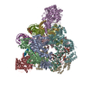
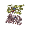
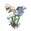
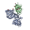
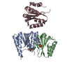
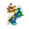
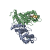
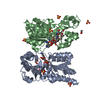
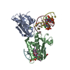
- PDB-4nik: Structure of human Gankyrin in complex to the single chain antibody F5 -

+
Open data


ID or keywords:

Loading...

-
Basic information

Entry
Database: PDB / ID: 4nik
TitleStructure of human Gankyrin in complex to the single chain antibody F5
Components
  • 26S proteasome non-ATPase regulatory subunit 10
  • Single-chain Fv fragment antibody
KeywordsOncoprotein/Immune System / beta-hairpin-alpha-hairpin repeat / Oncoprotein / 26S proteasome / Oncoprotein-Immune System complex
Function / homology
Function and homology information


proteasome regulatory particle assembly / positive regulation of cyclin-dependent protein serine/threonine kinase activity / Proteasome assembly / transcription factor binding / negative regulation of release of cytochrome c from mitochondria / negative regulation of NF-kappaB transcription factor activity / negative regulation of DNA damage response, signal transduction by p53 class mediator / negative regulation of MAPK cascade / proteasome complex / positive regulation of protein ubiquitination ...proteasome regulatory particle assembly / positive regulation of cyclin-dependent protein serine/threonine kinase activity / Proteasome assembly / transcription factor binding / negative regulation of release of cytochrome c from mitochondria / negative regulation of NF-kappaB transcription factor activity / negative regulation of DNA damage response, signal transduction by p53 class mediator / negative regulation of MAPK cascade / proteasome complex / positive regulation of protein ubiquitination / positive regulation of proteasomal ubiquitin-dependent protein catabolic process / microtubule cytoskeleton / positive regulation of cell growth / RNA polymerase II-specific DNA-binding transcription factor binding / cytoskeleton / cilium / apoptotic process / regulation of transcription by RNA polymerase II / negative regulation of apoptotic process / negative regulation of transcription by RNA polymerase II / nucleus / cytosol / cytoplasm
Similarity search - Function
: / Ankyrin repeat-containing domain / Ankyrin repeat / Ankyrin repeats (3 copies) / Ankyrin repeat profile. / Ankyrin repeat region circular profile. / ankyrin repeats / Ankyrin repeat / Ankyrin repeat-containing domain superfamily / Serine Threonine Protein Phosphatase 5, Tetratricopeptide repeat ...: / Ankyrin repeat-containing domain / Ankyrin repeat / Ankyrin repeats (3 copies) / Ankyrin repeat profile. / Ankyrin repeat region circular profile. / ankyrin repeats / Ankyrin repeat / Ankyrin repeat-containing domain superfamily / Serine Threonine Protein Phosphatase 5, Tetratricopeptide repeat / Alpha Horseshoe / Immunoglobulins / Immunoglobulin-like / Sandwich / Mainly Beta / Mainly Alpha
Similarity search - Domain/homology
26S proteasome non-ATPase regulatory subunit 10
Similarity search - Component
Biological speciesHomo sapiens (human)
MethodX-RAY DIFFRACTION / SYNCHROTRON / MOLECULAR REPLACEMENT / Resolution: 2.5 Å
AuthorsSato, Y. / Weiss, E. / Rochel, N.
CitationJournal: J.Mol.Biol. / Year: 2014
Title: Restricted diversity of antigen binding residues of antibodies revealed by computational alanine scanning of 227 antibody-antigen complexes
Authors: Robin, G. / Sato, Y. / Desplancq, D. / Rochel, N. / Weiss, E. / Martineau, P.
History
DepositionNov 6, 2013Deposition site: RCSB / Processing site: PDBJ
Revision 1.0Sep 10, 2014Provider: repository / Type: Initial release
Revision 1.1Oct 1, 2014Group: Database references / Source and taxonomy
Revision 1.2Sep 23, 2015Group: Database references
Revision 1.3Nov 8, 2023Group: Data collection / Database references / Refinement description
Category: chem_comp_atom / chem_comp_bond ...chem_comp_atom / chem_comp_bond / database_2 / pdbx_initial_refinement_model / struct_ref_seq_dif
Item: _database_2.pdbx_DOI / _database_2.pdbx_database_accession / _struct_ref_seq_dif.details
Revision 1.4Oct 16, 2024Group: Structure summary / Category: pdbx_entry_details / pdbx_modification_feature

-
Structure visualization

Structure viewerMolecule:
MolmilJmol/JSmol

Downloads & links

-
Assembly

Deposited unit
A: 26S proteasome non-ATPase regulatory subunit 10
B: Single-chain Fv fragment antibody


Theoretical massNumber of molelcules
Total (without water)51,7382
Polymers51,7382
Non-polymers00
Water7,710428
1


  • Idetical with deposited unit
  • defined by author&software
TypeNameSymmetry operationNumber
identity operation1_555x,y,z1
Buried area1680 Å2
ΔGint-7 kcal/mol
Surface area19470 Å2
MethodPISA
Unit cell
Length a, b, c (Å)135.578, 135.578, 68.655
Angle α, β, γ (deg.)90.00, 90.00, 120.00
Int Tables number154
Space group name H-MP3221
Components on special symmetry positions
IDModelComponents
11A-459-

HOH

-
Components

#1: Protein 26S proteasome non-ATPase regulatory subunit 10 / 26S proteasome regulatory subunit p28 / Gankyrin / p28(GANK)


Mass: 24873.326 Da / Num. of mol.: 1
Source method: isolated from a genetically manipulated source
Source: (gene. exp.) Homo sapiens (human) / Gene: PSMD10 / Production host: Escherichia coli (E. coli) / References: UniProt: O75832
#2: Antibody Single-chain Fv fragment antibody


Mass: 26864.672 Da / Num. of mol.: 1 / Source method: obtained synthetically / Details: semi synthetic scFv / Source: (synth.) Homo sapiens (human)
#3: Water ChemComp-HOH / water


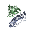
Mass: 18.015 Da / Num. of mol.: 428 / Source method: isolated from a natural source / Formula: H2O
Has protein modificationY

-
Experimental details

-
Experiment

ExperimentMethod: X-RAY DIFFRACTION / Number of used crystals: 1

-
Sample preparation

CrystalDensity Matthews: 3.52 Å3/Da / Density % sol: 65.06 %
Crystal growTemperature: 290 K / Method: vapor diffusion, sitting drop / pH: 7
Details: 8% PEG8000, 0.1M Hepes, pH 7.0, VAPOR DIFFUSION, SITTING DROP, temperature 290K

-
Data collection

DiffractionMean temperature: 100 K
Diffraction sourceSource: SYNCHROTRON / Site: ESRF / Beamline: ID29
DetectorType: ADSC QUANTUM 4r / Detector: CCD / Date: Jun 1, 2008
RadiationMonochromator: Si 111 CHANNEL / Protocol: SINGLE WAVELENGTH / Monochromatic (M) / Laue (L): M / Scattering type: x-ray
Radiation wavelengthRelative weight: 1
ReflectionResolution: 2.5→30 Å / Num. all: 25163 / Num. obs: 25163 / % possible obs: 98.9 % / Observed criterion σ(F): 2 / Observed criterion σ(I): 2 / Biso Wilson estimate: 29.98 Å2
Reflection shellResolution: 2.5→2.59 Å / % possible all: 97.9

-
Processing

Software
NameVersionClassification
ADSCQuantumdata collection
AMoREphasing
PHENIX(phenix.refine)refinement
HKL-2000data reduction
HKL-2000data scaling
RefinementMethod to determine structure: MOLECULAR REPLACEMENT
Starting model: 1UOH, 3KFU
Resolution: 2.5→29.633 Å / Occupancy max: 1 / Occupancy min: 0 / FOM work R set: 0.8739 / SU ML: 0.61 / σ(F): 1.34 / Phase error: 19.67 / Stereochemistry target values: ML
RfactorNum. reflection% reflectionSelection details
Rfree0.2078 1284 5.11 %random
Rwork0.1636 ---
all0.1658 25162 --
obs0.1658 25115 98.84 %-
Solvent computationShrinkage radii: 0.9 Å / VDW probe radii: 1.11 Å / Solvent model: FLAT BULK SOLVENT MODEL / Bsol: 66.531 Å2 / ksol: 0.318 e/Å3
Displacement parametersBiso max: 125.78 Å2 / Biso mean: 41.7502 Å2 / Biso min: 9.4 Å2
Baniso -1Baniso -2Baniso -3
1-2.9597 Å2-0 Å2-0 Å2
2--2.9597 Å2-0 Å2
3----5.9193 Å2
Refinement stepCycle: LAST / Resolution: 2.5→29.633 Å
ProteinNucleic acidLigandSolventTotal
Num. atoms3419 0 0 428 3847
LS refinement shell

Refine-ID: X-RAY DIFFRACTION / Total num. of bins used: 9

Resolution (Å)Rfactor RfreeNum. reflection RfreeRfactor RworkNum. reflection RworkNum. reflection all% reflection obs (%)
2.5003-2.60030.26631440.20982599274398
2.6003-2.71860.25171510.20022607275899
2.7186-2.86180.25011370.18492642277999
2.8618-3.04090.23481340.18942623275799
3.0409-3.27540.20591500.166526432793100
3.2754-3.60450.21541520.148226592811100
3.6045-4.12490.18571450.14382656280199
4.1249-5.19240.18521310.14132649278098
5.1924-29.63550.17191400.15362753289398
Refinement TLS params.Method: refined / Origin x: 4.0257 Å / Origin y: 55.9162 Å / Origin z: 29.7362 Å
111213212223313233
T0.084 Å20.0323 Å2-0.0138 Å2-0.061 Å20.0447 Å2--0.0728 Å2
L0.8452 °2-0.2557 °2-0.3265 °2-0.4999 °20.1369 °2--0.9138 °2
S-0.0107 Å °-0.1847 Å °-0.1543 Å °0.0184 Å °0.0045 Å °0.0588 Å °0.203 Å °0.1357 Å °0.034 Å °
Refinement TLS groupSelection details: ALL

+
About Yorodumi

-
News

-
Feb 9, 2022. New format data for meta-information of EMDB entries

New format data for meta-information of EMDB entries

  • Version 3 of the EMDB header file is now the official format.
  • The previous official version 1.9 will be removed from the archive.

Related info.:EMDB header

External links:wwPDB to switch to version 3 of the EMDB data model

-
Aug 12, 2020. Covid-19 info

Covid-19 info

URL: https://pdbj.org/emnavi/covid19.php

New page: Covid-19 featured information page in EM Navigator.

Related info.:Covid-19 info / Mar 5, 2020. Novel coronavirus structure data

+
Mar 5, 2020. Novel coronavirus structure data

Novel coronavirus structure data

Related info.:Yorodumi Speices / Aug 12, 2020. Covid-19 info

External links:COVID-19 featured content - PDBj / Molecule of the Month (242):Coronavirus Proteases

+
Jan 31, 2019. EMDB accession codes are about to change! (news from PDBe EMDB page)

EMDB accession codes are about to change! (news from PDBe EMDB page)

  • The allocation of 4 digits for EMDB accession codes will soon come to an end. Whilst these codes will remain in use, new EMDB accession codes will include an additional digit and will expand incrementally as the available range of codes is exhausted. The current 4-digit format prefixed with “EMD-” (i.e. EMD-XXXX) will advance to a 5-digit format (i.e. EMD-XXXXX), and so on. It is currently estimated that the 4-digit codes will be depleted around Spring 2019, at which point the 5-digit format will come into force.
  • The EM Navigator/Yorodumi systems omit the EMD- prefix.

Related info.:Q: What is EMD? / ID/Accession-code notation in Yorodumi/EM Navigator

External links:EMDB Accession Codes are Changing Soon! / Contact to PDBj

+
Jul 12, 2017. Major update of PDB

Major update of PDB

  • wwPDB released updated PDB data conforming to the new PDBx/mmCIF dictionary.
  • This is a major update changing the version number from 4 to 5, and with Remediation, in which all the entries are updated.
  • In this update, many items about electron microscopy experimental information are reorganized (e.g. em_software).
  • Now, EM Navigator and Yorodumi are based on the updated data.

External links:wwPDB Remediation / Enriched Model Files Conforming to OneDep Data Standards Now Available in the PDB FTP Archive

-
Yorodumi

Thousand views of thousand structures

  • Yorodumi is a browser for structure data from EMDB, PDB, SASBDB, etc.
  • This page is also the successor to EM Navigator detail page, and also detail information page/front-end page for Omokage search.
  • The word "yorodu" (or yorozu) is an old Japanese word meaning "ten thousand". "mi" (miru) is to see.

Related info.:EMDB / PDB / SASBDB / Comparison of 3 databanks / Yorodumi Search / Aug 31, 2016. New EM Navigator & Yorodumi / Yorodumi Papers / Jmol/JSmol / Function and homology information / Changes in new EM Navigator and Yorodumi

Read more