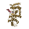
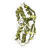
[English] 日本語
Yorodumi
- PDB-4gba: DCNL complex with N-terminally acetylated NEDD8 E2 peptide -

+
Open data


ID or keywords:

Loading...

-
Basic information

Entry
Database: PDB / ID: 4gba
TitleDCNL complex with N-terminally acetylated NEDD8 E2 peptide
Components
  • DCN1-like protein 3
  • NEDD8-conjugating enzyme UBE2F
Keywordsligase/peptide / E3 ligase / ligase-peptide complex
Function / homology
Function and homology information


NEDD8 conjugating enzyme activity / ubiquitin-like protein binding / cullin-RING-type E3 NEDD8 transferase / NEDD8 transferase activity / protein neddylation / ubiquitin conjugating enzyme binding / positive regulation of ubiquitin-protein transferase activity / NEDD8 ligase activity / response to UV-C / negative regulation of G1/S transition of mitotic cell cycle ...NEDD8 conjugating enzyme activity / ubiquitin-like protein binding / cullin-RING-type E3 NEDD8 transferase / NEDD8 transferase activity / protein neddylation / ubiquitin conjugating enzyme binding / positive regulation of ubiquitin-protein transferase activity / NEDD8 ligase activity / response to UV-C / negative regulation of G1/S transition of mitotic cell cycle / cullin family protein binding / ubiquitin ligase complex / post-translational protein modification / response to gamma radiation / negative regulation of cell growth / Antigen processing: Ubiquitination & Proteasome degradation / Neddylation / positive regulation of apoptotic process / perinuclear region of cytoplasm / ATP binding / nucleus / plasma membrane / cytosol
Similarity search - Function
Cullin, PONY binding domain / Potentiating neddylation domain / Defective-in-cullin neddylation protein / DCN1-like, PONY binding domain / Cullin binding / DCUN1 domain profile. / Ubiquitin-conjugating enzyme, active site / Ubiquitin-conjugating (UBC) active site signature. / Ubiquitin-conjugating enzyme E2, catalytic domain homologues / Ubiquitin-conjugating enzyme E2 ...Cullin, PONY binding domain / Potentiating neddylation domain / Defective-in-cullin neddylation protein / DCN1-like, PONY binding domain / Cullin binding / DCUN1 domain profile. / Ubiquitin-conjugating enzyme, active site / Ubiquitin-conjugating (UBC) active site signature. / Ubiquitin-conjugating enzyme E2, catalytic domain homologues / Ubiquitin-conjugating enzyme E2 / Ubiquitin-conjugating enzyme / Ubiquitin-conjugating (UBC) core domain profile. / Ubiquitin-conjugating enzyme/RWD-like / EF-hand / Recoverin; domain 1 / Orthogonal Bundle / Mainly Alpha
Similarity search - Domain/homology
DCN1-like protein 3 / NEDD8-conjugating enzyme UBE2F
Similarity search - Component
Biological speciesHomo sapiens (human)
MethodX-RAY DIFFRACTION / SYNCHROTRON / MOLECULAR REPLACEMENT / Resolution: 2.4 Å
AuthorsMonda, J.K. / Scott, D.C. / Miller, D.J. / Harper, J.W. / Bennett, E.J. / Schulman, B.A.
CitationJournal: Structure / Year: 2013
Title: Structural Conservation of Distinctive N-terminal Acetylation-Dependent Interactions across a Family of Mammalian NEDD8 Ligation Enzymes.
Authors: Monda, J.K. / Scott, D.C. / Miller, D.J. / Lydeard, J. / King, D. / Harper, J.W. / Bennett, E.J. / Schulman, B.A.
History
DepositionJul 26, 2012Deposition site: RCSB / Processing site: RCSB
Revision 1.0Nov 28, 2012Provider: repository / Type: Initial release
Revision 1.1Dec 19, 2012Group: Database references
Revision 1.2Jan 30, 2013Group: Database references

-
Structure visualization

Structure viewerMolecule:
MolmilJmol/JSmol

Downloads & links

-
Assembly

Deposited unit
A: DCN1-like protein 3
B: DCN1-like protein 3
F: NEDD8-conjugating enzyme UBE2F
G: NEDD8-conjugating enzyme UBE2F


Theoretical massNumber of molelcules
Total (without water)56,6524
Polymers56,6524
Non-polymers00
Water2,378132
1
A: DCN1-like protein 3
F: NEDD8-conjugating enzyme UBE2F


Theoretical massNumber of molelcules
Total (without water)28,3262
Polymers28,3262
Non-polymers00
Water362
TypeNameSymmetry operationNumber
identity operation1_555x,y,z1
Buried area1100 Å2
ΔGint-15 kcal/mol
Surface area10530 Å2
MethodPISA
2
B: DCN1-like protein 3
G: NEDD8-conjugating enzyme UBE2F


Theoretical massNumber of molelcules
Total (without water)28,3262
Polymers28,3262
Non-polymers00
Water362
TypeNameSymmetry operationNumber
identity operation1_555x,y,z1
Buried area1040 Å2
ΔGint-17 kcal/mol
Surface area11180 Å2
MethodPISA
Unit cell
Length a, b, c (Å)83.465, 44.578, 101.216
Angle α, β, γ (deg.)90.00, 103.30, 90.00
Int Tables number4
Space group name H-MP1211

-
Components

#1: Protein DCN1-like protein 3 / DCUN1 domain-containing protein 3 / Defective in cullin neddylation protein 1-like protein 3


Mass: 25671.791 Da / Num. of mol.: 2
Source method: isolated from a genetically manipulated source
Source: (gene. exp.) Homo sapiens (human) / Gene: DCUN1D3 / Production host: Escherichia coli (E. coli) / References: UniProt: Q8IWE4
#2: Protein/peptide NEDD8-conjugating enzyme UBE2F / NEDD8 carrier protein UBE2F / NEDD8 protein ligase UBE2F / NEDD8-conjugating enzyme 2 / Ubiquitin- ...NEDD8 carrier protein UBE2F / NEDD8 protein ligase UBE2F / NEDD8-conjugating enzyme 2 / Ubiquitin-conjugating enzyme E2 F


Mass: 2654.029 Da / Num. of mol.: 2 / Source method: obtained synthetically / Source: (synth.) Homo sapiens (human) / References: UniProt: Q969M7
#3: Water ChemComp-HOH / water / Water


Mass: 18.015 Da / Num. of mol.: 132 / Source method: isolated from a natural source / Formula: H2O

-
Experimental details

-
Experiment

ExperimentMethod: X-RAY DIFFRACTION / Number of used crystals: 1

-
Sample preparation

CrystalDensity Matthews: 3.23 Å3/Da / Density % sol: 61.97 %
Crystal growTemperature: 277 K / Method: vapor diffusion, hanging drop / pH: 7
Details: crystals in 2.28M sodium malonate, seeded into 2.05M sodium malonate, pH 7.0, VAPOR DIFFUSION, HANGING DROP, temperature 277K

-
Data collection

DiffractionMean temperature: 77 K
Diffraction sourceSource: SYNCHROTRON / Site: ALS / Beamline: 8.2.2 / Wavelength: 0.9791 Å
DetectorType: ADSC QUANTUM 315r / Detector: CCD / Date: Jan 14, 2011
RadiationMonochromator: Double crystal, Si(111) / Protocol: SINGLE WAVELENGTH / Monochromatic (M) / Laue (L): M / Scattering type: x-ray
Radiation wavelengthWavelength: 0.9791 Å / Relative weight: 1
ReflectionResolution: 2.4→29.1 Å / Num. all: 28832 / Num. obs: 28812 / % possible obs: 99.7 % / Observed criterion σ(F): 0 / Observed criterion σ(I): -3
Reflection shell
Resolution (Å)Diffraction-ID% possible all
2.4-2.49197.9
2.49-2.59198.8
2.59-2.7199.9
2.7-2.851100
2.85-3.021100
3.02-3.261100
3.26-3.581100
3.58-4.11100
4.1-5.161100
5.16-301100

-
Processing

Software
NameVersionClassification
BOSdata collection
PHASERphasing
PHENIX(phenix.refine: 1.7.1_743)refinement
HKL-2000data reduction
HKL-2000data scaling
RefinementMethod to determine structure: MOLECULAR REPLACEMENT / Resolution: 2.4→29.085 Å / SU ML: 0.54 / σ(F): 1.34 / Phase error: 23.89 / Stereochemistry target values: ML
RfactorNum. reflection% reflectionSelection details
Rfree0.2273 1466 5.09 %random
Rwork0.1939 ---
obs0.1956 28812 99.44 %-
all-28832 --
Solvent computationShrinkage radii: 0.9 Å / VDW probe radii: 1.11 Å / Solvent model: FLAT BULK SOLVENT MODEL / Bsol: 46.838 Å2 / ksol: 0.403 e/Å3
Displacement parameters
Baniso -1Baniso -2Baniso -3
1-12.1497 Å2-0 Å2-9.9209 Å2
2---10.8347 Å20 Å2
3----0.8215 Å2
Refinement stepCycle: LAST / Resolution: 2.4→29.085 Å
ProteinNucleic acidLigandSolventTotal
Num. atoms3403 0 0 132 3535
Refine LS restraints
Refine-IDTypeDev idealNumber
X-RAY DIFFRACTIONf_bond_d0.0083481
X-RAY DIFFRACTIONf_angle_d1.0324701
X-RAY DIFFRACTIONf_dihedral_angle_d15.2851263
X-RAY DIFFRACTIONf_chiral_restr0.069499
X-RAY DIFFRACTIONf_plane_restr0.004613
LS refinement shell
Resolution (Å)Rfactor RfreeNum. reflection RfreeRfactor RworkNum. reflection RworkRefine-ID% reflection obs (%)
2.4-2.48280.311460.26742608X-RAY DIFFRACTION96
2.4828-2.58220.31131330.25532703X-RAY DIFFRACTION99
2.5822-2.69960.29441330.2362737X-RAY DIFFRACTION100
2.6996-2.84180.27081580.21362703X-RAY DIFFRACTION100
2.8418-3.01970.28691450.20782717X-RAY DIFFRACTION100
3.0197-3.25260.22321620.18912722X-RAY DIFFRACTION100
3.2526-3.57940.21691440.1872744X-RAY DIFFRACTION100
3.5794-4.09610.20211530.16712771X-RAY DIFFRACTION100
4.0961-5.1560.18121310.15932780X-RAY DIFFRACTION100
5.156-29.08710.20941610.20472861X-RAY DIFFRACTION100

+
About Yorodumi

-
News

-
Feb 9, 2022. New format data for meta-information of EMDB entries

New format data for meta-information of EMDB entries

  • Version 3 of the EMDB header file is now the official format.
  • The previous official version 1.9 will be removed from the archive.

Related info.:EMDB header

External links:wwPDB to switch to version 3 of the EMDB data model

-
Aug 12, 2020. Covid-19 info

Covid-19 info

URL: https://pdbj.org/emnavi/covid19.php

New page: Covid-19 featured information page in EM Navigator.

Related info.:Covid-19 info / Mar 5, 2020. Novel coronavirus structure data

+
Mar 5, 2020. Novel coronavirus structure data

Novel coronavirus structure data

Related info.:Yorodumi Speices / Aug 12, 2020. Covid-19 info

External links:COVID-19 featured content - PDBj / Molecule of the Month (242):Coronavirus Proteases

+
Jan 31, 2019. EMDB accession codes are about to change! (news from PDBe EMDB page)

EMDB accession codes are about to change! (news from PDBe EMDB page)

  • The allocation of 4 digits for EMDB accession codes will soon come to an end. Whilst these codes will remain in use, new EMDB accession codes will include an additional digit and will expand incrementally as the available range of codes is exhausted. The current 4-digit format prefixed with “EMD-” (i.e. EMD-XXXX) will advance to a 5-digit format (i.e. EMD-XXXXX), and so on. It is currently estimated that the 4-digit codes will be depleted around Spring 2019, at which point the 5-digit format will come into force.
  • The EM Navigator/Yorodumi systems omit the EMD- prefix.

Related info.:Q: What is EMD? / ID/Accession-code notation in Yorodumi/EM Navigator

External links:EMDB Accession Codes are Changing Soon! / Contact to PDBj

+
Jul 12, 2017. Major update of PDB

Major update of PDB

  • wwPDB released updated PDB data conforming to the new PDBx/mmCIF dictionary.
  • This is a major update changing the version number from 4 to 5, and with Remediation, in which all the entries are updated.
  • In this update, many items about electron microscopy experimental information are reorganized (e.g. em_software).
  • Now, EM Navigator and Yorodumi are based on the updated data.

External links:wwPDB Remediation / Enriched Model Files Conforming to OneDep Data Standards Now Available in the PDB FTP Archive

-
Yorodumi

Thousand views of thousand structures

  • Yorodumi is a browser for structure data from EMDB, PDB, SASBDB, etc.
  • This page is also the successor to EM Navigator detail page, and also detail information page/front-end page for Omokage search.
  • The word "yorodu" (or yorozu) is an old Japanese word meaning "ten thousand". "mi" (miru) is to see.

Related info.:EMDB / PDB / SASBDB / Comparison of 3 databanks / Yorodumi Search / Aug 31, 2016. New EM Navigator & Yorodumi / Yorodumi Papers / Jmol/JSmol / Function and homology information / Changes in new EM Navigator and Yorodumi

Read more