 Movie
Movie Controller


+ Open data

- Basic information
| Entry | Database: PDB / ID: 2fha | ||||||
|---|---|---|---|---|---|---|---|
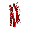
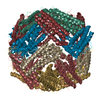
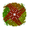
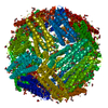
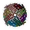
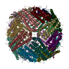
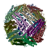
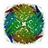
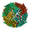
| Title | HUMAN H CHAIN FERRITIN | ||||||
 Components Components | FERRITIN | ||||||
 Keywords Keywords | IRON STORAGE | ||||||
| Function / homology |  Function and homology information Function and homology informationiron ion sequestering activity / ferritin complex / Scavenging by Class A Receptors / Golgi Associated Vesicle Biogenesis / ferroxidase / negative regulation of ferroptosis / autolysosome / ferroxidase activity / negative regulation of fibroblast proliferation / ferric iron binding ...iron ion sequestering activity / ferritin complex / Scavenging by Class A Receptors / Golgi Associated Vesicle Biogenesis / ferroxidase / negative regulation of ferroptosis / autolysosome / ferroxidase activity / negative regulation of fibroblast proliferation / ferric iron binding / autophagosome / iron ion transport / ferrous iron binding / Iron uptake and transport / tertiary granule lumen / ficolin-1-rich granule lumen / intracellular iron ion homeostasis / immune response / iron ion binding / negative regulation of cell population proliferation / Neutrophil degranulation / extracellular exosome / extracellular region / identical protein binding / nucleus / cytoplasm / cytosol Similarity search - Function | ||||||
| Biological species |  Homo sapiens (human) Homo sapiens (human) | ||||||
| Method |  X-RAY DIFFRACTION / SYNCHROTRON / MOLECULAR REPLACEMENT / Resolution: 1.9 Å X-RAY DIFFRACTION / SYNCHROTRON / MOLECULAR REPLACEMENT / Resolution: 1.9 Å | ||||||
 Authors Authors | Hempstead, P.D. / Artymiuk, P.J. / Harrison, P.M. | ||||||
 Citation Citation | Journal: J.Mol.Biol. / Year: 1997 Title: Comparison of the three-dimensional structures of recombinant human H and horse L ferritins at high resolution. Authors: Hempstead, P.D. / Yewdall, S.J. / Fernie, A.R. / Lawson, D.M. / Artymiuk, P.J. / Rice, D.W. / Ford, G.C. / Harrison, P.M. #1: Journal: Nature / Year: 1991Title: Solving the Structure of Human H Ferritin by Genetically Engineering Intermolecular Crystal Contacts Authors: Lawson, D.M. / Artymiuk, P.J. / Yewdall, S.J. / Smith, J.M. / Livingstone, J.C. / Treffry, A. / Luzzago, A. / Levi, S. / Arosio, P. / Cesareni, G. / Thomas, C.D. / Shaw, W.V. / Harrison, P.M. | ||||||
| History |
|
- Structure visualization
| Structure viewer | Molecule: MolmilJmol/JSmol |
|---|
- Downloads & links
-Download
| PDBx/mmCIF format | 2fha.cif.gz | 50 KB | Display | PDBx/mmCIF format |
|---|---|---|---|---|
| PDB format | pdb2fha.ent.gz | 36.7 KB | Display | PDB format |
| PDBx/mmJSON format | 2fha.json.gz | Tree view | PDBx/mmJSON format | |
| Others |  Other downloads Other downloads |
-Validation report
| Arichive directory | https://data.pdbj.org/pub/pdb/validation_reports/fh/2fhaftp://data.pdbj.org/pub/pdb/validation_reports/fh/2fha | HTTPS FTP |
|---|
-Related structure data
| Related structure data | 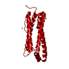 1aewC 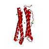 1fhaS S: Starting model for refinement C: citing same article ( |
|---|---|
| Similar structure data |


-Links
 PDBj
PDBj


- Assembly
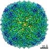
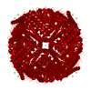
| Deposited unit | 
| ||||||||
|---|---|---|---|---|---|---|---|---|---|
| 1 | x 24
| ||||||||
| Unit cell |
| ||||||||
| Components on special symmetry positions |
|
-Components
| #1: Protein | Mass: 21254.605 Da / Num. of mol.: 1 / Mutation: K86Q Source method: isolated from a genetically manipulated source Source: (gene. exp.) Homo sapiens (human)Description: SEE D.M.LAWSON,P.J.ARTYMIUK,S.J.YEWDALL, J.M.A.SMITH,J.C.LIVINGSTONE,A.TREFFRY,A.LUZZAGO,S.LEVI, P.AROSIO,G.CESARINI,C.D.THOMAS,W.V.SHAW,P.M.HARRISON, NATURE 1991, 349, P541 Production host:  | ||
|---|---|---|---|
| #2: Chemical |   Mass: 40.078 Da / Num. of mol.: 2 / Source method: obtained synthetically / Formula: Ca Mass: 40.078 Da / Num. of mol.: 2 / Source method: obtained synthetically / Formula: Ca#3: Water | ChemComp-HOH / |  Mass: 18.015 Da / Num. of mol.: 59 / Source method: isolated from a natural source / Formula: H2O Mass: 18.015 Da / Num. of mol.: 59 / Source method: isolated from a natural source / Formula: H2O |
-Experimental details
-Experiment
| Experiment | Method: X-RAY DIFFRACTION / Number of used crystals: 1 |
|---|
- Sample preparation
| Crystal | Density Matthews: 3.09 Å3/Da / Density % sol: 60.23 % | ||||||||||||||||||||||||||||||||||||
|---|---|---|---|---|---|---|---|---|---|---|---|---|---|---|---|---|---|---|---|---|---|---|---|---|---|---|---|---|---|---|---|---|---|---|---|---|---|
| Crystal grow | pH: 7.5 Details: PROTEIN WAS CRYSTALLIZED FROM 0.08% CACL2 / 15% MPD IN 50 MM HEPES BUFFER PH 7.5 | ||||||||||||||||||||||||||||||||||||
| Crystal grow | *PLUS Method: vapor diffusion, hanging drop | ||||||||||||||||||||||||||||||||||||
| Components of the solutions | *PLUS
|
-Data collection
| Diffraction | Mean temperature: 293 K |
|---|---|
| Diffraction source | Source: SYNCHROTRON / Site: SRS  / Beamline: PX9.6 / Wavelength: 0.915 / Beamline: PX9.6 / Wavelength: 0.915 |
| Detector | Type: KODAK / Detector: FILM / Date: Nov 14, 1988 / Details: MIRRORS |
| Radiation | Monochromator: SI(111) / Monochromatic (M) / Laue (L): M / Scattering type: x-ray |
| Radiation wavelength | Wavelength: 0.915 Å / Relative weight: 1 |
| Reflection | Resolution: 1.9→20 Å / Num. obs: 20238 / % possible obs: 94 % / Observed criterion σ(I): 0 / Redundancy: 7.2 % / Biso Wilson estimate: 18.2 Å2 / Rmerge(I) obs: 0.076 / Rsym value: 0.076 / Net I/σ(I): 9 |
| Reflection shell | Resolution: 1.9→2 Å / Redundancy: 5.3 % / Rmerge(I) obs: 0.567 / Mean I/σ(I) obs: 2.4 / Rsym value: 0.567 / % possible all: 90 |
| Reflection shell | *PLUS % possible obs: 90 % |
- Processing
| Software |
| ||||||||||||||||||||||||||||||||||||||||||||||||||
|---|---|---|---|---|---|---|---|---|---|---|---|---|---|---|---|---|---|---|---|---|---|---|---|---|---|---|---|---|---|---|---|---|---|---|---|---|---|---|---|---|---|---|---|---|---|---|---|---|---|---|---|
| Refinement | Method to determine structure: MOLECULAR REPLACEMENT Starting model: PDB ENTRY 1FHA Resolution: 1.9→20 Å / σ(F): 0 / Stereochemistry target values: TNT PROTGEO
| ||||||||||||||||||||||||||||||||||||||||||||||||||
| Solvent computation | Solvent model: DEFAULT / Bsol: 300 Å2 / ksol: 0.85 e/Å3 | ||||||||||||||||||||||||||||||||||||||||||||||||||
| Refinement step | Cycle: LAST / Resolution: 1.9→20 Å
| ||||||||||||||||||||||||||||||||||||||||||||||||||
| Refine LS restraints |
| ||||||||||||||||||||||||||||||||||||||||||||||||||
| Software | *PLUS Name: TNT / Version: 5D / Classification: refinement | ||||||||||||||||||||||||||||||||||||||||||||||||||
| Refinement | *PLUS | ||||||||||||||||||||||||||||||||||||||||||||||||||
| Solvent computation | *PLUS | ||||||||||||||||||||||||||||||||||||||||||||||||||
| Displacement parameters | *PLUS | ||||||||||||||||||||||||||||||||||||||||||||||||||
| Refine LS restraints | *PLUS
|
