 Movie
Movie Controller


+ Open data

- Basic information
| Entry | Database: PDB / ID: 1aew | ||||||
|---|---|---|---|---|---|---|---|
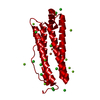
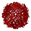
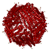
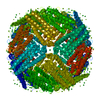
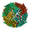
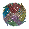
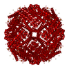
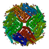
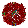
| Title | L-CHAIN HORSE APOFERRITIN | ||||||
 Components Components | FERRITIN | ||||||
 Keywords Keywords | IRON STORAGE / MULTIGENE FAMILY | ||||||
| Function / homology |  Function and homology information Function and homology informationferritin complex / autolysosome / ferric iron binding / autophagosome / iron ion transport / ferrous iron binding / cytoplasmic vesicle / intracellular iron ion homeostasis / iron ion binding / cytoplasm Similarity search - Function | ||||||
| Biological species |  | ||||||
| Method |  X-RAY DIFFRACTION / SYNCHROTRON / MOLECULAR REPLACEMENT / Resolution: 1.95 Å X-RAY DIFFRACTION / SYNCHROTRON / MOLECULAR REPLACEMENT / Resolution: 1.95 Å | ||||||
 Authors Authors | Hempstead, P.D. / Yewdall, S.J. / Lawson, D.M. / Harrison, P.M. / Artymiuk, P.J. | ||||||
 Citation Citation | Journal: J.Mol.Biol. / Year: 1997 Title: Comparison of the three-dimensional structures of recombinant human H and horse L ferritins at high resolution. Authors: Hempstead, P.D. / Yewdall, S.J. / Fernie, A.R. / Lawson, D.M. / Artymiuk, P.J. / Rice, D.W. / Ford, G.C. / Harrison, P.M. | ||||||
| History |
|
- Structure visualization
| Structure viewer | Molecule: MolmilJmol/JSmol |
|---|
- Downloads & links
-Download
| PDBx/mmCIF format | 1aew.cif.gz | 50 KB | Display | PDBx/mmCIF format |
|---|---|---|---|---|
| PDB format | pdb1aew.ent.gz | 36.7 KB | Display | PDB format |
| PDBx/mmJSON format | 1aew.json.gz | Tree view | PDBx/mmJSON format | |
| Others |  Other downloads Other downloads |
-Validation report
| Arichive directory | https://data.pdbj.org/pub/pdb/validation_reports/ae/1aewftp://data.pdbj.org/pub/pdb/validation_reports/ae/1aew | HTTPS FTP |
|---|
-Related structure data


-Links
 PDBj
PDBj


- Assembly
| Deposited unit | 
| ||||||||
|---|---|---|---|---|---|---|---|---|---|
| 1 | x 24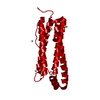
| ||||||||
| Unit cell |
| ||||||||
| Components on special symmetry positions |
|
-Components
| #1: Protein | Mass: 19856.385 Da / Num. of mol.: 1 / Fragment: L CHAIN Source method: isolated from a genetically manipulated source Source: (gene. exp.) Description: SEE TAKEDA ET AL. 1993 BIOCHIM. BIOPHYS. ACTA. 1174, 218-220. Organ: SPLEEN / Plasmid: PTZ18U / Production host:  | ||
|---|---|---|---|
| #2: Chemical | ChemComp-CD /   Mass: 112.411 Da / Num. of mol.: 6 / Source method: obtained synthetically / Formula: Cd Mass: 112.411 Da / Num. of mol.: 6 / Source method: obtained synthetically / Formula: Cd#3: Water | ChemComp-HOH / |  Mass: 18.015 Da / Num. of mol.: 76 / Source method: isolated from a natural source / Formula: H2O Mass: 18.015 Da / Num. of mol.: 76 / Source method: isolated from a natural source / Formula: H2O |
-Experimental details
-Experiment
| Experiment | Method: X-RAY DIFFRACTION / Number of used crystals: 1 |
|---|
- Sample preparation
| Crystal | Density Matthews: 3.27 Å3/Da / Density % sol: 62.36 % | |||||||||||||||||||||
|---|---|---|---|---|---|---|---|---|---|---|---|---|---|---|---|---|---|---|---|---|---|---|
| Crystal grow | Method: vapor diffusion, sitting drop / pH: 7 Details: CRYSTALLIZED BY SITTING DROP: 15 LAMBDA PROTEIN + 1-2 LAMBDA 2% CDSO4, pH 7.0, vapor diffusion - sitting drop | |||||||||||||||||||||
| Crystal grow | *PLUS Method: vapor diffusion, sitting drop | |||||||||||||||||||||
| Components of the solutions | *PLUS
|
-Data collection
| Diffraction | Mean temperature: 290 K |
|---|---|
| Diffraction source | Source: SYNCHROTRON / Site: SRS  / Beamline: PX9.5 / Wavelength: 0.92 / Beamline: PX9.5 / Wavelength: 0.92 |
| Detector | Type: MARRESEARCH / Detector: IMAGE PLATE / Date: Feb 1, 1993 / Details: MIRRORS |
| Radiation | Monochromator: SI(111) / Monochromatic (M) / Laue (L): M / Scattering type: x-ray |
| Radiation wavelength | Wavelength: 0.92 Å / Relative weight: 1 |
| Reflection | Resolution: 1.95→20 Å / Num. obs: 19915 / % possible obs: 100 % / Observed criterion σ(I): 0 / Redundancy: 7.6 % / Biso Wilson estimate: 15.9 Å2 / Rmerge(I) obs: 0.03 / Net I/σ(I): 17.6 |
| Reflection shell | Resolution: 1.95→2 Å / Redundancy: 4.2 % / Rmerge(I) obs: 0.12 / Mean I/σ(I) obs: 6.3 / % possible all: 97.5 |
| Reflection shell | *PLUS % possible obs: 97.5 % |
- Processing
| Software |
| ||||||||||||||||||||||||||||||||||||||||||||||||||
|---|---|---|---|---|---|---|---|---|---|---|---|---|---|---|---|---|---|---|---|---|---|---|---|---|---|---|---|---|---|---|---|---|---|---|---|---|---|---|---|---|---|---|---|---|---|---|---|---|---|---|---|
| Refinement | Method to determine structure: MOLECULAR REPLACEMENT Starting model: HORSE SPLEEN L FERRITIN. Resolution: 1.95→20 Å / σ(F): 0 / Stereochemistry target values: TNT PROTGEO
| ||||||||||||||||||||||||||||||||||||||||||||||||||
| Solvent computation | Solvent model: DEFAULT / Bsol: 412 Å2 / ksol: 0.85 e/Å3 | ||||||||||||||||||||||||||||||||||||||||||||||||||
| Refinement step | Cycle: LAST / Resolution: 1.95→20 Å
| ||||||||||||||||||||||||||||||||||||||||||||||||||
| Refine LS restraints |
| ||||||||||||||||||||||||||||||||||||||||||||||||||
| Software | *PLUS Name: TNT / Version: 5D / Classification: refinement | ||||||||||||||||||||||||||||||||||||||||||||||||||
| Refinement | *PLUS | ||||||||||||||||||||||||||||||||||||||||||||||||||
| Solvent computation | *PLUS | ||||||||||||||||||||||||||||||||||||||||||||||||||
| Displacement parameters | *PLUS | ||||||||||||||||||||||||||||||||||||||||||||||||||
| Refine LS restraints | *PLUS
|
