+ Open data
- Basic information
| Entry | Database: PDB / ID: 4hug | ||||||
|---|---|---|---|---|---|---|---|
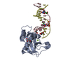
| Title | Structure of 5-chlorouracil modified A:U base pairs | ||||||
 Components Components |
| ||||||
 Keywords Keywords | Hydrolase/DNA / 5-chloro-2'-deoxyuridine / W-C base pair / Wobble base pair / double helix / Watson-Crick base pairing pattern / Hydrolase-DNA complex | ||||||
| Function / homology |  Function and homology information Function and homology informationDNA replication, removal of RNA primer / ribonuclease H / RNA-DNA hybrid ribonuclease activity / nucleic acid binding / metal ion binding / cytoplasm Similarity search - Function | ||||||
| Biological species |  Bacillus halodurans (bacteria) Bacillus halodurans (bacteria) | ||||||
| Method |  X-RAY DIFFRACTION / X-RAY DIFFRACTION /  SYNCHROTRON / SYNCHROTRON /  MOLECULAR REPLACEMENT / Resolution: 1.64 Å MOLECULAR REPLACEMENT / Resolution: 1.64 Å | ||||||
 Authors Authors | Patra, A. / Egli, M. | ||||||
 Citation Citation |  Journal: Nucleic Acids Res. / Year: 2013 Journal: Nucleic Acids Res. / Year: 2013Title: Structure, stability and function of 5-chlorouracil modified A:U and G:U base pairs. Authors: Patra, A. / Harp, J. / Pallan, P.S. / Zhao, L. / Abramov, M. / Herdewijn, P. / Egli, M. | ||||||
| History |
|
- Structure visualization
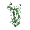
| Structure viewer | Molecule:  Molmil Molmil Jmol/JSmol Jmol/JSmol |
|---|
- Downloads & links
- Download
| PDBx/mmCIF format |  4hug.cif.gz 4hug.cif.gz | 105.9 KB | Display |  PDBx/mmCIF format PDBx/mmCIF format |
|---|---|---|---|---|
| PDB format |  pdb4hug.ent.gz pdb4hug.ent.gz | 77.2 KB | Display |  PDB format PDB format |
| PDBx/mmJSON format |  4hug.json.gz 4hug.json.gz | Tree view |  PDBx/mmJSON format PDBx/mmJSON format | |
| Others |  Other downloads Other downloads |
-Validation report
| Arichive directory |  https://data.pdbj.org/pub/pdb/validation_reports/hu/4hug https://data.pdbj.org/pub/pdb/validation_reports/hu/4hug ftp://data.pdbj.org/pub/pdb/validation_reports/hu/4hug ftp://data.pdbj.org/pub/pdb/validation_reports/hu/4hug | HTTPS FTP |
|---|
-Related structure data
| Related structure data |  4htuC  4hueC  4hufC  3d0pS C: citing same article ( S: Starting model for refinement |
|---|---|
| Similar structure data |
- Links
Links
- Assembly
| Deposited unit | 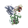
| ||||||||
|---|---|---|---|---|---|---|---|---|---|
| 1 | 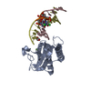
| ||||||||
| 2 | 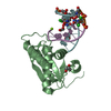
| ||||||||
| Unit cell |
|
- Components
-Protein / DNA chain , 2 types, 6 molecules ABCDEF
| #1: Protein | Mass: 15429.371 Da / Num. of mol.: 2 / Mutation: D132N Source method: isolated from a genetically manipulated source Source: (gene. exp.)  Bacillus halodurans (bacteria) Bacillus halodurans (bacteria)Strain: ATCC BAA-125 / DSM 18197 / FERM 7344 / JCM 9153 / C-125 Gene: rnhA, BH0863 / Production host:  #2: DNA chain | Mass: 3704.230 Da / Num. of mol.: 4 / Source method: obtained synthetically / Details: Dickerson Dodecamer DNA, ClU7/8 |
|---|
-Non-polymers , 4 types, 434 molecules 


| #3: Chemical | | #4: Chemical | #5: Chemical | #6: Water | ChemComp-HOH / | |
|---|
-Experimental details
-Experiment
| Experiment | Method:  X-RAY DIFFRACTION / Number of used crystals: 1 X-RAY DIFFRACTION / Number of used crystals: 1 |
|---|
- Sample preparation
| Crystal | Density Matthews: 2.63 Å3/Da / Density % sol: 53.16 % |
|---|---|
| Crystal grow | Temperature: 277 K / pH: 6.5 Details: 0.2 M magnesium acetate, 0.1 M sodium cacodylate and 20% (w/v) PEG 8000 , pH 6.5, VAPOR DIFFUSION, SITTING DROP, temperature 277K |
-Data collection
| Diffraction | Mean temperature: 100 K |
|---|---|
| Diffraction source | Source:  SYNCHROTRON / Site: SYNCHROTRON / Site:  APS APS  / Beamline: 21-ID-F / Wavelength: 0.97872 / Beamline: 21-ID-F / Wavelength: 0.97872 |
| Detector | Type: MARMOSAIC 225 mm CCD / Detector: CCD / Date: Jul 11, 2011 |
| Radiation | Monochromator: C(111) / Protocol: SINGLE WAVELENGTH / Monochromatic (M) / Laue (L): M / Scattering type: x-ray |
| Radiation wavelength | Wavelength: 0.97872 Å / Relative weight: 1 |
| Reflection | Resolution: 1.64→50 Å / Num. obs: 59379 / % possible obs: 99.7 % / Observed criterion σ(I): -3 / Redundancy: 8.5 % |
| Reflection shell | Resolution: 1.64→1.67 Å / Rmerge(I) obs: 0.674 / % possible all: 99.3 |
- Processing
| Software |
| ||||||||||||||||||||||||||||||||||||||||||||||||||||||||||||||||||||||||||||||||||||||||||||||||||||||||||||||||||||||||||||||||||||||||||||||||||||||||||
|---|---|---|---|---|---|---|---|---|---|---|---|---|---|---|---|---|---|---|---|---|---|---|---|---|---|---|---|---|---|---|---|---|---|---|---|---|---|---|---|---|---|---|---|---|---|---|---|---|---|---|---|---|---|---|---|---|---|---|---|---|---|---|---|---|---|---|---|---|---|---|---|---|---|---|---|---|---|---|---|---|---|---|---|---|---|---|---|---|---|---|---|---|---|---|---|---|---|---|---|---|---|---|---|---|---|---|---|---|---|---|---|---|---|---|---|---|---|---|---|---|---|---|---|---|---|---|---|---|---|---|---|---|---|---|---|---|---|---|---|---|---|---|---|---|---|---|---|---|---|---|---|---|---|---|---|
| Refinement | Method to determine structure:  MOLECULAR REPLACEMENT MOLECULAR REPLACEMENTStarting model: 3D0P Resolution: 1.64→43.228 Å / SU ML: 0.16 / Isotropic thermal model: Isotropic / σ(F): 1.35 / Phase error: 20.95 / Stereochemistry target values: ML
| ||||||||||||||||||||||||||||||||||||||||||||||||||||||||||||||||||||||||||||||||||||||||||||||||||||||||||||||||||||||||||||||||||||||||||||||||||||||||||
| Solvent computation | Shrinkage radii: 0.9 Å / VDW probe radii: 1.1 Å / Solvent model: FLAT BULK SOLVENT MODEL | ||||||||||||||||||||||||||||||||||||||||||||||||||||||||||||||||||||||||||||||||||||||||||||||||||||||||||||||||||||||||||||||||||||||||||||||||||||||||||
| Refinement step | Cycle: LAST / Resolution: 1.64→43.228 Å
| ||||||||||||||||||||||||||||||||||||||||||||||||||||||||||||||||||||||||||||||||||||||||||||||||||||||||||||||||||||||||||||||||||||||||||||||||||||||||||
| Refine LS restraints |
| ||||||||||||||||||||||||||||||||||||||||||||||||||||||||||||||||||||||||||||||||||||||||||||||||||||||||||||||||||||||||||||||||||||||||||||||||||||||||||
| LS refinement shell |
|
 Movie
Movie Controller





 PDBj
PDBj


