 Movie
Movie Controller

[English] 日本語
 Yorodumi
- PDB-3d0p: Insights into RNA/DNA hybrid recognition and processing by RNase ... -
+ Open data

- Basic information
| Entry | Database: PDB / ID: 3d0p | ||||||
|---|---|---|---|---|---|---|---|
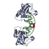
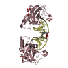
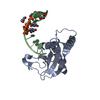
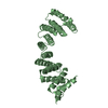
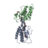
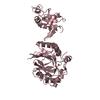
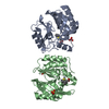
| Title | Insights into RNA/DNA hybrid recognition and processing by RNase H from the crystal structure of a non-specific enzyme-dsDNA complex | ||||||
 Components Components |
| ||||||
 Keywords Keywords | hydrolase/DNA / RNase H-DNA complex / A-form / B-form / metal ions / protein-DNA complex / Cytoplasm / Endonuclease / Hydrolase / Magnesium / Manganese / Metal-binding / Nuclease / hydrolase-DNA COMPLEX | ||||||
| Function / homology |  Function and homology information Function and homology informationDNA replication, removal of RNA primer / ribonuclease H / RNA-DNA hybrid ribonuclease activity / nucleic acid binding / metal ion binding / cytoplasm Similarity search - Function | ||||||
| Method |  X-RAY DIFFRACTION / SYNCHROTRON / MOLECULAR REPLACEMENT / Resolution: 1.8 Å X-RAY DIFFRACTION / SYNCHROTRON / MOLECULAR REPLACEMENT / Resolution: 1.8 Å | ||||||
 Authors Authors | Pallan, P.S. / Egli, M. | ||||||
 Citation Citation | Journal: Cell Cycle / Year: 2008 Title: Insights into RNA/DNA hybrid recognition and processing by RNase H from the crystal structure of a non-specific enzyme-dsDNA complex. Authors: Pallan, P.S. / Egli, M. | ||||||
| History |
|
- Structure visualization
| Structure viewer | Molecule: MolmilJmol/JSmol |
|---|
- Downloads & links
-Download
| PDBx/mmCIF format | 3d0p.cif.gz | 85.8 KB | Display | PDBx/mmCIF format |
|---|---|---|---|---|
| PDB format | pdb3d0p.ent.gz | 61.5 KB | Display | PDB format |
| PDBx/mmJSON format | 3d0p.json.gz | Tree view | PDBx/mmJSON format | |
| Others |  Other downloads Other downloads |
-Validation report
| Arichive directory | https://data.pdbj.org/pub/pdb/validation_reports/d0/3d0pftp://data.pdbj.org/pub/pdb/validation_reports/d0/3d0p | HTTPS FTP |
|---|
-Related structure data
| Related structure data |  1zbiS S: Starting model for refinement |
|---|---|
| Similar structure data |


-Links
 PDBj
PDBj


- Assembly
| Deposited unit | 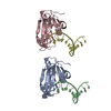
| ||||||||
|---|---|---|---|---|---|---|---|---|---|
| 1 | 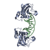
| ||||||||
| 2 | 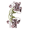
| ||||||||
| Unit cell |
| ||||||||
| Details | The second part of the biological assembly is generated by the two fold axis of Chain identifier A: -X, Y, -Z -1.000000 0.000000 0.000000 0.000000 1.000000 0.000000 0.000000 0.000000 -1.000000 / The second part of the biological assembly is generated by the two fold axis of Chain identifier B: -X, Y, -Z -1.000000 0.000000 0.000000 0.000000 1.000000 0.000000 0.000000 0.000000 -1.000000 |
-Components
| #1: Protein | Mass: 15429.371 Da / Num. of mol.: 2 / Mutation: D132N / Source method: obtained synthetically / Details: Bacillus halodurans RNase H, D132N mutant / References: UniProt: Q9KEI9, ribonuclease H #2: DNA chain | Mass: 3663.392 Da / Num. of mol.: 2 / Source method: obtained synthetically / Details: Synthetic DNA #3: Chemical | ChemComp-NA / |   Mass: 22.990 Da / Num. of mol.: 1 / Source method: obtained synthetically / Formula: Na Mass: 22.990 Da / Num. of mol.: 1 / Source method: obtained synthetically / Formula: Na#4: Water | ChemComp-HOH / |  Mass: 18.015 Da / Num. of mol.: 166 / Source method: isolated from a natural source / Formula: H2O Mass: 18.015 Da / Num. of mol.: 166 / Source method: isolated from a natural source / Formula: H2O |
|---|
-Experimental details
-Experiment
| Experiment | Method: X-RAY DIFFRACTION / Number of used crystals: 1 |
|---|
- Sample preparation
| Crystal | Density Matthews: 2.79 Å3/Da / Density % sol: 55.94 % | ||||||||||||||||||||
|---|---|---|---|---|---|---|---|---|---|---|---|---|---|---|---|---|---|---|---|---|---|
| Crystal grow | Temperature: 291 K / Method: vapor diffusion, sitting drop / pH: 4.6 Details: 0.1 M sodium acetate, 8 % (w/v) PEG 4000, pH 4.6, VAPOR DIFFUSION, SITTING DROP, temperature 291K | ||||||||||||||||||||
| Components of the solutions |
|
-Data collection
| Diffraction | Mean temperature: 100 K |
|---|---|
| Diffraction source | Source: SYNCHROTRON / Site: APS  / Beamline: 21-ID-F / Wavelength: 0.9785 Å / Beamline: 21-ID-F / Wavelength: 0.9785 Å |
| Detector | Type: MARMOSAIC 225 mm CCD / Detector: CCD / Date: Oct 21, 2007 |
| Radiation | Monochromator: C(111) / Protocol: SINGLE WAVELENGTH / Monochromatic (M) / Laue (L): M / Scattering type: x-ray |
| Radiation wavelength | Wavelength: 0.9785 Å / Relative weight: 1 |
| Reflection | Resolution: 1.8→50 Å / Num. all: 38816 / Num. obs: 37768 / % possible obs: 97.3 % / Observed criterion σ(F): 0 / Observed criterion σ(I): 0 / Redundancy: 7.7 % / Rmerge(I) obs: 0.061 / Net I/σ(I): 12.4 |
| Reflection shell | Resolution: 1.8→1.86 Å / Redundancy: 7.3 % / Rmerge(I) obs: 0.248 / Mean I/σ(I) obs: 12.4 / Num. unique all: 3643 / % possible all: 94.7 |
- Processing
| Software |
| |||||||||||||||||||||||||||||||||||||||||||||||||||||||||||||||||
|---|---|---|---|---|---|---|---|---|---|---|---|---|---|---|---|---|---|---|---|---|---|---|---|---|---|---|---|---|---|---|---|---|---|---|---|---|---|---|---|---|---|---|---|---|---|---|---|---|---|---|---|---|---|---|---|---|---|---|---|---|---|---|---|---|---|---|
| Refinement | Method to determine structure: MOLECULAR REPLACEMENT Starting model: PDB ENTRY 1ZBI, using one protein molecule. Resolution: 1.8→41.59 Å / Cor.coef. Fo:Fc: 0.961 / Cor.coef. Fo:Fc free: 0.951 / SU B: 5.491 / SU ML: 0.086 / Cross valid method: THROUGHOUT / ESU R: 0.133 / ESU R Free: 0.124 / Stereochemistry target values: MAXIMUM LIKELIHOOD / Details: HYDROGENS HAVE BEEN ADDED IN THE RIDING POSITIONS
| |||||||||||||||||||||||||||||||||||||||||||||||||||||||||||||||||
| Solvent computation | Ion probe radii: 0.8 Å / Shrinkage radii: 0.8 Å / VDW probe radii: 1.2 Å / Solvent model: MASK | |||||||||||||||||||||||||||||||||||||||||||||||||||||||||||||||||
| Displacement parameters | Biso mean: 31.474 Å2
| |||||||||||||||||||||||||||||||||||||||||||||||||||||||||||||||||
| Refinement step | Cycle: LAST / Resolution: 1.8→41.59 Å
| |||||||||||||||||||||||||||||||||||||||||||||||||||||||||||||||||
| Refine LS restraints |
| |||||||||||||||||||||||||||||||||||||||||||||||||||||||||||||||||
| LS refinement shell | Resolution: 1.803→1.849 Å / Total num. of bins used: 20
| |||||||||||||||||||||||||||||||||||||||||||||||||||||||||||||||||
| Refinement TLS params. | Method: refined / Origin x: 32.4421 Å / Origin y: 1.845 Å / Origin z: 18.6056 Å
| |||||||||||||||||||||||||||||||||||||||||||||||||||||||||||||||||
| Refinement TLS group |
|
