 Movie
Movie Controller

[English] 日本語
 Yorodumi
- PDB-3ey1: A Conformational Transition in the Structure of a 2'-Thiomethyl-M... -
+ Open data

- Basic information
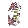
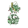
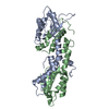
| Entry | Database: PDB / ID: 3ey1 | ||||||
|---|---|---|---|---|---|---|---|
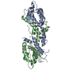
| Title | A Conformational Transition in the Structure of a 2'-Thiomethyl-Modified DNA Visualized at High Resolution | ||||||
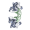
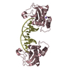
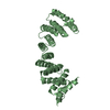
 Components Components |
| ||||||
 Keywords Keywords | HYDROLASE/DNA / RNase H-DNA COMPLEX / PROTEIN-DNA COMPLEX / ENDONUCLEASE / 2'-THIOMETHYL URIDINE RIBONUCLEIC ACID / Hydrolase / Magnesium / Manganese / Metal-binding / Nuclease / HYDROLASE-DNA COMPLEX | ||||||
| Function / homology |  Function and homology information Function and homology informationDNA replication, removal of RNA primer / ribonuclease H / RNA-DNA hybrid ribonuclease activity / nucleic acid binding / metal ion binding / cytoplasm Similarity search - Function | ||||||
| Biological species |  Bacillus halodurans (bacteria) Bacillus halodurans (bacteria) | ||||||
| Method |  X-RAY DIFFRACTION / SYNCHROTRON / MOLECULAR REPLACEMENT / Resolution: 1.6 Å X-RAY DIFFRACTION / SYNCHROTRON / MOLECULAR REPLACEMENT / Resolution: 1.6 Å | ||||||
 Authors Authors | Egli, M. / Pallan, P.S. | ||||||
 Citation Citation | Journal: Chem.Commun.(Camb.) / Year: 2009 Title: A conformational transition in the structure of a 2'-thiomethyl-modified DNA visualized at high resolution. Authors: Pallan, P.S. / Prakash, T.P. / Li, F. / Eoff, R.L. / Manoharan, M. / Egli, M. | ||||||
| History |
|
- Structure visualization
| Structure viewer | Molecule: MolmilJmol/JSmol |
|---|
- Downloads & links
-Download
| PDBx/mmCIF format | 3ey1.cif.gz | 88.1 KB | Display | PDBx/mmCIF format |
|---|---|---|---|---|
| PDB format | pdb3ey1.ent.gz | 62.2 KB | Display | PDB format |
| PDBx/mmJSON format | 3ey1.json.gz | Tree view | PDBx/mmJSON format | |
| Others |  Other downloads Other downloads |
-Validation report
| Arichive directory | https://data.pdbj.org/pub/pdb/validation_reports/ey/3ey1ftp://data.pdbj.org/pub/pdb/validation_reports/ey/3ey1 | HTTPS FTP |
|---|
-Related structure data
| Related structure data |  3ey2C 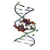 3ey3C  3d0pS C: citing same article ( S: Starting model for refinement |
|---|---|
| Similar structure data |


-Links
 PDBj
PDBj


- Assembly
| Deposited unit | 
| ||||||||
|---|---|---|---|---|---|---|---|---|---|
| 1 | 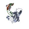
| ||||||||
| Unit cell |
|
-Components
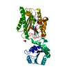
| #1: Protein | Mass: 15916.004 Da / Num. of mol.: 1 / Fragment: RNase H domain, UNP residues 59-196 / Mutation: D132N Source method: isolated from a genetically manipulated source Source: (gene. exp.) Bacillus halodurans (bacteria) / Gene: rnhA, BH0863 / References: UniProt: Q9KEI9, ribonuclease H | ||
|---|---|---|---|
| #2: DNA chain | Mass: 3727.522 Da / Num. of mol.: 1 / Source method: obtained synthetically Details: Chemically synthesized modified dodecamer DNA (oligonucleotide) | ||
| #3: Chemical |   Mass: 92.094 Da / Num. of mol.: 3 / Source method: obtained synthetically / Formula: C3H8O3 Mass: 92.094 Da / Num. of mol.: 3 / Source method: obtained synthetically / Formula: C3H8O3#4: Water | ChemComp-HOH / |  Mass: 18.015 Da / Num. of mol.: 103 / Source method: isolated from a natural source / Formula: H2O Mass: 18.015 Da / Num. of mol.: 103 / Source method: isolated from a natural source / Formula: H2O |
-Experimental details
-Experiment
| Experiment | Method: X-RAY DIFFRACTION / Number of used crystals: 1 |
|---|
- Sample preparation
| Crystal | Density Matthews: 2.62 Å3/Da / Density % sol: 53.07 % | ||||||||||||||||||||||||||||
|---|---|---|---|---|---|---|---|---|---|---|---|---|---|---|---|---|---|---|---|---|---|---|---|---|---|---|---|---|---|
| Crystal grow | Temperature: 291 K / Method: vapor diffusion, sitting drop / pH: 4.6 Details: 0.1 M Sodium Acetate (pH 4.6), 0.2 M ammonium sulphate, 25% PEG 4000., VAPOR DIFFUSION, SITTING DROP, temperature 291K | ||||||||||||||||||||||||||||
| Components of the solutions |
|
-Data collection
| Diffraction | Mean temperature: 120 K |
|---|---|
| Diffraction source | Source: SYNCHROTRON / Site: APS  / Beamline: 21-ID-F / Wavelength: 0.9785 Å / Beamline: 21-ID-F / Wavelength: 0.9785 Å |
| Detector | Type: MARMOSAIC 225 mm CCD / Detector: CCD / Date: Oct 20, 2007 |
| Radiation | Protocol: SINGLE WAVELENGTH / Monochromatic (M) / Laue (L): M / Scattering type: x-ray |
| Radiation wavelength | Wavelength: 0.9785 Å / Relative weight: 1 |
| Reflection | Resolution: 1.6→50 Å / Num. all: 26658 / Num. obs: 26499 / % possible obs: 99.4 % / Observed criterion σ(I): 0 / Redundancy: 5.8 % / Rmerge(I) obs: 0.052 / Net I/σ(I): 363.5 |
| Reflection shell | Resolution: 1.6→1.66 Å / Redundancy: 5 % / Rmerge(I) obs: 0.29 / Mean I/σ(I) obs: 9.9 / Num. unique all: 2562 / % possible all: 96.9 |
- Processing
| Software |
| |||||||||||||||||||||||||
|---|---|---|---|---|---|---|---|---|---|---|---|---|---|---|---|---|---|---|---|---|---|---|---|---|---|---|
| Refinement | Method to determine structure: MOLECULAR REPLACEMENT Starting model: PDB ENTRY 3D0P, USING THE PROTEIN MOLECULE ALONE. Resolution: 1.6→50 Å / Cross valid method: THROUGHOUT / σ(I): 0
| |||||||||||||||||||||||||
| Refinement step | Cycle: LAST / Resolution: 1.6→50 Å
| |||||||||||||||||||||||||
| Refine LS restraints |
|
