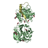
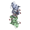
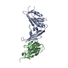
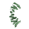
- PDB-3aaf: Structure of WRN RQC domain bound to double-stranded DNA -
+
Open data
ID or keywords:
Loading...
-
Basic information
Entry
Database: PDB / ID: 3aaf
Title
Structure of WRN RQC domain bound to double-stranded DNA
Components
DNA (5'-D(*AP*CP*CP*CP*TP*AP*AP*TP*TP*AP*GP*GP*GP*T)-3')
Werner syndrome ATP-dependent helicase
Keywords
DNA BINDING PROTEIN/DNA / HELIX-TURN-HELIX / WINGED-HELIX / PROTEIN-DNA COMPLEX / DNA-binding / Helicase / DNA BINDING PROTEIN-DNA complex
Function / homology
Function and homology information
3'-flap-structured DNA binding / positive regulation of strand invasion / positive regulation of hydrolase activity / forked DNA-dependent helicase activity / telomeric G-quadruplex DNA binding / 8-hydroxy-2'-deoxyguanosine DNA binding / telomeric D-loop binding / regulation of growth rate / DNA geometric change / telomere maintenance via semi-conservative replication ...3'-flap-structured DNA binding / positive regulation of strand invasion / positive regulation of hydrolase activity / forked DNA-dependent helicase activity / telomeric G-quadruplex DNA binding / 8-hydroxy-2'-deoxyguanosine DNA binding / telomeric D-loop binding / regulation of growth rate / DNA geometric change / telomere maintenance via semi-conservative replication / Y-form DNA binding / telomeric D-loop disassembly / four-way junction helicase activity / t-circle formation / G-quadruplex DNA binding / bubble DNA binding / MutLalpha complex binding / Impaired BRCA2 binding to PALB2 / protein localization to nucleolus / Processive synthesis on the C-strand of the telomere / response to UV-C / Removal of the Flap Intermediate from the C-strand / exonuclease activity / HDR through Single Strand Annealing (SSA) / DNA metabolic process / Homologous DNA Pairing and Strand Exchange / Defective homologous recombination repair (HRR) due to BRCA1 loss of function / Defective HDR through Homologous Recombination Repair (HRR) due to PALB2 loss of BRCA1 binding function / Defective HDR through Homologous Recombination Repair (HRR) due to PALB2 loss of BRCA2/RAD51/RAD51C binding function / Resolution of D-loop Structures through Synthesis-Dependent Strand Annealing (SDSA) / DNA synthesis involved in DNA repair / Resolution of D-loop Structures through Holliday Junction Intermediates / 3'-5' DNA helicase activity / Impaired BRCA2 binding to RAD51 / DNA 3'-5' helicase / replication fork processing / replicative senescence / Presynaptic phase of homologous DNA pairing and strand exchange / mismatch repair / SUMOylation of DNA damage response and repair proteins / four-way junction DNA binding / 3'-5' exonuclease activity / telomere maintenance / cellular response to starvation / replication fork / DNA helicase activity / determination of adult lifespan / cellular response to gamma radiation / base-excision repair / G2/M DNA damage checkpoint / double-strand break repair via homologous recombination / HDR through Homologous Recombination (HRR) / cellular senescence / manganese ion binding / double-strand break repair / chromosome / Processing of DNA double-strand break ends / response to oxidative stress / Regulation of TP53 Activity through Phosphorylation / Hydrolases; Acting on ester bonds / DNA replication / chromosome, telomeric region / nuclear speck / DNA damage response / centrosome / protein-containing complex binding / nucleolus / magnesium ion binding / protein homodimerization activity / ATP hydrolysis activity / DNA binding / nucleoplasm / ATP binding / nucleus / cytoplasm Similarity search - Function
A: Werner syndrome ATP-dependent helicase B: Werner syndrome ATP-dependent helicase C: DNA (5'-D(*AP*CP*CP*CP*TP*AP*AP*TP*TP*AP*GP*GP*GP*T)-3') D: DNA (5'-D(*AP*CP*CP*CP*TP*AP*AP*TP*TP*AP*GP*GP*GP*T)-3') hetero molecules
In the structure databanks used in Yorodumi, some data are registered as the other names, "COVID-19 virus" and "2019-nCoV". Here are the details of the virus and the list of structure data.
Jan 31, 2019. EMDB accession codes are about to change! (news from PDBe EMDB page)
EMDB accession codes are about to change! (news from PDBe EMDB page)
The allocation of 4 digits for EMDB accession codes will soon come to an end. Whilst these codes will remain in use, new EMDB accession codes will include an additional digit and will expand incrementally as the available range of codes is exhausted. The current 4-digit format prefixed with “EMD-” (i.e. EMD-XXXX) will advance to a 5-digit format (i.e. EMD-XXXXX), and so on. It is currently estimated that the 4-digit codes will be depleted around Spring 2019, at which point the 5-digit format will come into force.
The EM Navigator/Yorodumi systems omit the EMD- prefix.
Related info.:Q: What is EMD? / ID/Accession-code notation in Yorodumi/EM Navigator
Yorodumi is a browser for structure data from EMDB, PDB, SASBDB, etc.
This page is also the successor to EM Navigator detail page, and also detail information page/front-end page for Omokage search.
The word "yorodu" (or yorozu) is an old Japanese word meaning "ten thousand". "mi" (miru) is to see.
Related info.:EMDB / PDB / SASBDB / Comparison of 3 databanks / Yorodumi Search / Aug 31, 2016. New EM Navigator & Yorodumi / Yorodumi Papers / Jmol/JSmol / Function and homology information / Changes in new EM Navigator and Yorodumi
 Movie
Movie Controller


 Open data
 Basic information
 Components
 Keywords
 Function and homology information
 Homo sapiens (human)
 X-RAY DIFFRACTION /
  Authors
 Citation
 Structure visualization
 Downloads & links
 Other downloads
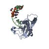


 PDBj
PDBj


 Assembly


 Mass: 59.044 Da / Num. of mol.: 2 / Source method: obtained synthetically / Formula: C2H3O2
 Mass: 18.015 Da / Num. of mol.: 338 / Source method: isolated from a natural source / Formula: H2O
 Sample preparation
 Processing