 Movie
Movie Controller


+ Open data

- Basic information
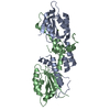
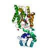
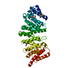
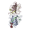
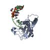
| Entry | Database: PDB / ID: 6k18 | ||||||
|---|---|---|---|---|---|---|---|
| Title | Crystal structure of EXD2 exonuclease domain soaked in Mn | ||||||
 Components Components | Exonuclease 3'-5' domain-containing protein 2 | ||||||
 Keywords Keywords | HYDROLASE / 3'-5' exonuclease / DnaQ family | ||||||
| Function / homology |  Function and homology information Function and homology informationexodeoxyribonuclease I / nucleic acid metabolic process / DNA double-strand break processing / single-stranded DNA 3'-5' DNA exonuclease activity / 3'-5'-DNA exonuclease activity / intermediate filament cytoskeleton / replication fork processing / site of DNA damage / 3'-5' exonuclease activity / double-strand break repair via homologous recombination ...exodeoxyribonuclease I / nucleic acid metabolic process / DNA double-strand break processing / single-stranded DNA 3'-5' DNA exonuclease activity / 3'-5'-DNA exonuclease activity / intermediate filament cytoskeleton / replication fork processing / site of DNA damage / 3'-5' exonuclease activity / double-strand break repair via homologous recombination / manganese ion binding / double-strand break repair / Hydrolases; Acting on ester bonds; Exoribonucleases producing 5'-phosphomonoesters / 3'-5'-RNA exonuclease activity / nucleic acid binding / mitochondrial outer membrane / mitochondrial matrix / magnesium ion binding / protein homodimerization activity / mitochondrion / nucleus / cytoplasm Similarity search - Function | ||||||
| Biological species |  Homo sapiens (human) Homo sapiens (human) | ||||||
| Method |  X-RAY DIFFRACTION / SYNCHROTRON / MOLECULAR REPLACEMENT / Resolution: 2.303 Å X-RAY DIFFRACTION / SYNCHROTRON / MOLECULAR REPLACEMENT / Resolution: 2.303 Å | ||||||
 Authors Authors | Park, J. / Lee, C. | ||||||
 Citation Citation | Journal: Nucleic Acids Res. / Year: 2019 Title: The structure of human EXD2 reveals a chimeric 3' to 5' exonuclease domain that discriminates substrates via metal coordination. Authors: Park, J. / Lee, S.Y. / Jeong, H. / Kang, M.G. / Van Haute, L. / Minczuk, M. / Seo, J.K. / Jun, Y. / Myung, K. / Rhee, H.W. / Lee, C. | ||||||
| History |
|
- Structure visualization
| Structure viewer | Molecule: MolmilJmol/JSmol |
|---|
- Downloads & links
-Download
| PDBx/mmCIF format | 6k18.cif.gz | 95.9 KB | Display | PDBx/mmCIF format |
|---|---|---|---|---|
| PDB format | pdb6k18.ent.gz | 70.8 KB | Display | PDB format |
| PDBx/mmJSON format | 6k18.json.gz | Tree view | PDBx/mmJSON format | |
| Others |  Other downloads Other downloads |
-Validation report
| Arichive directory | https://data.pdbj.org/pub/pdb/validation_reports/k1/6k18ftp://data.pdbj.org/pub/pdb/validation_reports/k1/6k18 | HTTPS FTP |
|---|
-Related structure data
| Related structure data | 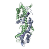 6k17SC 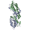 6k19C 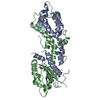 6k1aC 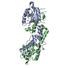 6k1bC  6k1cC 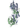 6k1dC 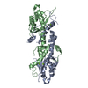 6k1eC S: Starting model for refinement C: citing same article ( |
|---|---|
| Similar structure data |


-Links
 PDBj
PDBj
- Assembly
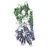
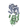
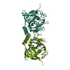
| Deposited unit | 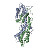
| ||||||||
|---|---|---|---|---|---|---|---|---|---|
| 1 |
| ||||||||
| Unit cell |
|
-Components
| #1: Protein | Mass: 24396.246 Da / Num. of mol.: 2 Source method: isolated from a genetically manipulated source Source: (gene. exp.) Homo sapiens (human) / Gene: EXD2, C14orf114, EXDL2 / Production host:  #2: Chemical |   Mass: 54.938 Da / Num. of mol.: 2 / Source method: obtained synthetically / Formula: Mn / Feature type: SUBJECT OF INVESTIGATION Mass: 54.938 Da / Num. of mol.: 2 / Source method: obtained synthetically / Formula: Mn / Feature type: SUBJECT OF INVESTIGATION#3: Water | ChemComp-HOH / |  Mass: 18.015 Da / Num. of mol.: 223 / Source method: isolated from a natural source / Formula: H2O Mass: 18.015 Da / Num. of mol.: 223 / Source method: isolated from a natural source / Formula: H2O |
|---|
-Experimental details
-Experiment
| Experiment | Method: X-RAY DIFFRACTION / Number of used crystals: 1 |
|---|
- Sample preparation
| Crystal | Density Matthews: 2.3 Å3/Da / Density % sol: 46.6 % |
|---|---|
| Crystal grow | Temperature: 277 K / Method: vapor diffusion, hanging drop Details: PEG 8000, 2-(N-morpholino)ethanesulfonic acid, Calcium acetate, Dithiothreitol, Manganese chloride |
-Data collection
| Diffraction | Mean temperature: 100 K / Serial crystal experiment: N |
|---|---|
| Diffraction source | Source: SYNCHROTRON / Site: PAL/PLS  / Beamline: 5C (4A) / Wavelength: 0.9795 Å / Beamline: 5C (4A) / Wavelength: 0.9795 Å |
| Detector | Type: DECTRIS PILATUS3 6M / Detector: PIXEL / Date: Oct 31, 2016 |
| Radiation | Protocol: SINGLE WAVELENGTH / Monochromatic (M) / Laue (L): M / Scattering type: x-ray |
| Radiation wavelength | Wavelength: 0.9795 Å / Relative weight: 1 |
| Reflection | Resolution: 2.3→50 Å / Num. obs: 20317 / % possible obs: 98.3 % / Redundancy: 5.2 % / Rmerge(I) obs: 0.103 / Net I/σ(I): 12.35 |
| Reflection shell | Resolution: 2.3→2.34 Å / Rmerge(I) obs: 0.348 / Num. unique obs: 939 |
- Processing
| Software |
| ||||||||||||||||||||||||||||||||||||||||||||||||||||||||
|---|---|---|---|---|---|---|---|---|---|---|---|---|---|---|---|---|---|---|---|---|---|---|---|---|---|---|---|---|---|---|---|---|---|---|---|---|---|---|---|---|---|---|---|---|---|---|---|---|---|---|---|---|---|---|---|---|---|
| Refinement | Method to determine structure: MOLECULAR REPLACEMENT Starting model: 6K17 Resolution: 2.303→43.609 Å / SU ML: 0.28 / Cross valid method: FREE R-VALUE / σ(F): 1.44 / Phase error: 25.28
| ||||||||||||||||||||||||||||||||||||||||||||||||||||||||
| Solvent computation | Shrinkage radii: 0.9 Å / VDW probe radii: 1.11 Å | ||||||||||||||||||||||||||||||||||||||||||||||||||||||||
| Refinement step | Cycle: LAST / Resolution: 2.303→43.609 Å
| ||||||||||||||||||||||||||||||||||||||||||||||||||||||||
| Refine LS restraints |
| ||||||||||||||||||||||||||||||||||||||||||||||||||||||||
| LS refinement shell |
|
