Entry Database : PDB / ID : 3wg5Title 1510-N membrane-bound stomatin-specific protease K138A mutant in complex with a substrate peptide under heat treatment 441aa long hypothetical nfeD protein PH1511 stomatin Keywords / / / / / Function / homology Function Domain/homology Component
/ / / / / / / / / / / / / / / / / / / / / / / / / / / / / / / / / / / Biological species Pyrococcus horikoshii (archaea)Pyrococcus horikoshii OT3 (archaea)Method / / / Resolution : 2.4 Å Authors Yokoyama, H. / Fujii, S. / Matsui, I. History Deposition Jul 26, 2013 Deposition site / Processing site Revision 1.0 Oct 23, 2013 Provider / Type Revision 1.1 Jan 1, 2020 Group / Category / struct_ref_seq_difItem _citation.country / _citation.journal_abbrev ... _citation.country / _citation.journal_abbrev / _citation.journal_id_CSD / _citation.journal_id_ISSN / _citation.pdbx_database_id_PubMed / _citation.title / _struct_ref_seq_dif.details Revision 1.2 Nov 8, 2023 Group Data collection / Database references ... Data collection / Database references / Derived calculations / Refinement description Category chem_comp_atom / chem_comp_bond ... chem_comp_atom / chem_comp_bond / database_2 / pdbx_initial_refinement_model / struct_site Item _database_2.pdbx_DOI / _database_2.pdbx_database_accession ... _database_2.pdbx_DOI / _database_2.pdbx_database_accession / _struct_site.pdbx_auth_asym_id / _struct_site.pdbx_auth_comp_id / _struct_site.pdbx_auth_seq_id
Show all Show less
 Movie
Movie Controller

 Yorodumi
 Open data
 Basic information
 Components
 Keywords
 Function and homology information
 Pyrococcus horikoshii (archaea)
 X-RAY DIFFRACTION /
  Authors
 Citation
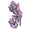
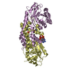
 Structure visualization
 Downloads & links
 Other downloads
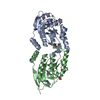


 PDBj
PDBj


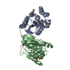
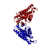
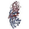
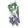
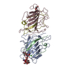
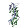
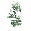
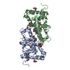
 Assembly
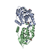


 Mass: 92.094 Da / Num. of mol.: 2 / Source method: obtained synthetically / Formula: C3H8O3
 Mass: 69.085 Da / Num. of mol.: 3 / Source method: obtained synthetically / Formula: C3H5N2
 Mass: 35.453 Da / Num. of mol.: 1 / Source method: obtained synthetically / Formula: Cl
 Sample preparation
 / Beamline: BL26B1 / Wavelength: 1 Å
 Processing