 Movie
Movie Controller

[English] 日本語
 Yorodumi
- PDB-3bpp: 1510-N membrane protease K138A mutant specific for a stomatin hom... -
+ Open data

- Basic information
| Entry | Database: PDB / ID: 3bpp | ||||||
|---|---|---|---|---|---|---|---|
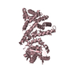
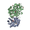
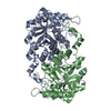
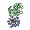
| Title | 1510-N membrane protease K138A mutant specific for a stomatin homolog from Pyrococcus horikoshii | ||||||
 Components Components | 1510-N membrane protease | ||||||
 Keywords Keywords | HYDROLASE / membrane protease / specific for a stomatin homolog / archaea / Pyrococcus / thermostable / catalytic dyad | ||||||
| Function / homology |  Function and homology information Function and homology informationHydrolases; Acting on peptide bonds (peptidases); Serine endopeptidases / serine-type peptidase activity / proteolysis / membrane Similarity search - Function | ||||||
| Biological species |   Pyrococcus horikoshii (archaea) Pyrococcus horikoshii (archaea) | ||||||
| Method |  X-RAY DIFFRACTION / SYNCHROTRON / MOLECULAR REPLACEMENT / Resolution: 2.3 Å X-RAY DIFFRACTION / SYNCHROTRON / MOLECULAR REPLACEMENT / Resolution: 2.3 Å | ||||||
 Authors Authors | Yokoyama, H. / Hamamatsu, S. / Fujii, S. / Matsui, I. | ||||||
 Citation Citation | Journal: J.SYNCHROTRON RADIAT. / Year: 2008 Title: Novel dimer structure of a membrane-bound protease with a catalytic Ser-Lys dyad and its linkage to stomatin Authors: Yokoyama, H. / Hamamatsu, S. / Fujii, S. / Matsui, I. #1: Journal: J.Mol.Biol. / Year: 2006Title: Molecular structure of a novel membrane protease specific for a stomatin homolog from the hyperthermophilic archaeon Pyrococcus horikoshii Authors: Yokoyama, H. / Matsui, E. / Akiba, T. / Harata, K. / Matsui, I. | ||||||
| History |
|
- Structure visualization
| Structure viewer | Molecule: MolmilJmol/JSmol |
|---|
- Downloads & links
-Download
| PDBx/mmCIF format | 3bpp.cif.gz | 56.5 KB | Display | PDBx/mmCIF format |
|---|---|---|---|---|
| PDB format | pdb3bpp.ent.gz | 40.3 KB | Display | PDB format |
| PDBx/mmJSON format | 3bpp.json.gz | Tree view | PDBx/mmJSON format | |
| Others |  Other downloads Other downloads |
-Validation report
| Arichive directory | https://data.pdbj.org/pub/pdb/validation_reports/bp/3bppftp://data.pdbj.org/pub/pdb/validation_reports/bp/3bpp | HTTPS FTP |
|---|
-Related structure data
| Related structure data |  2deoS S: Starting model for refinement |
|---|---|
| Similar structure data |


-Links
 PDBj
PDBj- Assembly
| Deposited unit | 
| ||||||||
|---|---|---|---|---|---|---|---|---|---|
| 1 | 
| ||||||||
| 2 | 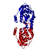
| ||||||||
| 3 |
| ||||||||
| Unit cell |
|
-Components
| #1: Protein | Mass: 25368.025 Da / Num. of mol.: 1 / Fragment: Residues 16-236 / Mutation: K138A Source method: isolated from a genetically manipulated source Source: (gene. exp.) Pyrococcus horikoshii (archaea) / Gene: PH1510 / Plasmid: pET21b / Production host:  |
|---|---|
| #2: Water | ChemComp-HOH /  Mass: 18.015 Da / Num. of mol.: 63 / Source method: isolated from a natural source / Formula: H2O Mass: 18.015 Da / Num. of mol.: 63 / Source method: isolated from a natural source / Formula: H2O |
-Experimental details
-Experiment
| Experiment | Method: X-RAY DIFFRACTION / Number of used crystals: 1 |
|---|
- Sample preparation
| Crystal | Density Matthews: 3.59 Å3/Da / Density % sol: 65.72 % |
|---|---|
| Crystal grow | Temperature: 293 K / Method: vapor diffusion, hanging drop / pH: 4.5 Details: 18% PEG4000, 0.1M magnesium chloride, 0.1M sodium acetate, pH 4.5, VAPOR DIFFUSION, HANGING DROP, temperature 293K |
-Data collection
| Diffraction | Mean temperature: 100 K |
|---|---|
| Diffraction source | Source: SYNCHROTRON / Site: Photon Factory  / Beamline: BL-6A / Wavelength: 1 Å / Beamline: BL-6A / Wavelength: 1 Å |
| Detector | Type: ADSC QUANTUM 4 / Detector: CCD / Date: Mar 17, 2006 / Details: mirror |
| Radiation | Monochromator: Si(111) / Protocol: SINGLE WAVELENGTH / Monochromatic (M) / Laue (L): M / Scattering type: x-ray |
| Radiation wavelength | Wavelength: 1 Å / Relative weight: 1 |
| Reflection | Resolution: 2.3→20 Å / Num. obs: 16792 / % possible obs: 99 % / Redundancy: 14.1 % / Biso Wilson estimate: 45.7 Å2 / Rmerge(I) obs: 0.049 / Net I/σ(I): 71.8 |
| Reflection shell | Resolution: 2.3→2.38 Å / Rmerge(I) obs: 0.33 / Mean I/σ(I) obs: 11.9 / % possible all: 100 |
- Processing
| Software |
| ||||||||||||||||||||||||||||||||||||||||||||||||||||||||||||||||||||||||||||||||||||||||||
|---|---|---|---|---|---|---|---|---|---|---|---|---|---|---|---|---|---|---|---|---|---|---|---|---|---|---|---|---|---|---|---|---|---|---|---|---|---|---|---|---|---|---|---|---|---|---|---|---|---|---|---|---|---|---|---|---|---|---|---|---|---|---|---|---|---|---|---|---|---|---|---|---|---|---|---|---|---|---|---|---|---|---|---|---|---|---|---|---|---|---|---|
| Refinement | Method to determine structure: MOLECULAR REPLACEMENT Starting model: PDB ENTRY 2DEO Resolution: 2.3→19.51 Å / Cor.coef. Fo:Fc: 0.933 / Cor.coef. Fo:Fc free: 0.882 / SU B: 8.011 / SU ML: 0.194 / Cross valid method: THROUGHOUT / ESU R: 0.259 / ESU R Free: 0.25 / Stereochemistry target values: MAXIMUM LIKELIHOOD
| ||||||||||||||||||||||||||||||||||||||||||||||||||||||||||||||||||||||||||||||||||||||||||
| Solvent computation | Ion probe radii: 0.8 Å / Shrinkage radii: 0.8 Å / VDW probe radii: 1.4 Å / Solvent model: MASK | ||||||||||||||||||||||||||||||||||||||||||||||||||||||||||||||||||||||||||||||||||||||||||
| Displacement parameters | Biso mean: 47.985 Å2 | ||||||||||||||||||||||||||||||||||||||||||||||||||||||||||||||||||||||||||||||||||||||||||
| Refine analyze |
| ||||||||||||||||||||||||||||||||||||||||||||||||||||||||||||||||||||||||||||||||||||||||||
| Refinement step | Cycle: LAST / Resolution: 2.3→19.51 Å
| ||||||||||||||||||||||||||||||||||||||||||||||||||||||||||||||||||||||||||||||||||||||||||
| Refine LS restraints |
| ||||||||||||||||||||||||||||||||||||||||||||||||||||||||||||||||||||||||||||||||||||||||||
| LS refinement shell | Resolution: 2.297→2.356 Å / Total num. of bins used: 20
|
