 Movie
Movie Controller

[English] 日本語
 Yorodumi
- PDB-1k3u: CRYSTAL STRUCTURE OF WILD-TYPE TRYPTOPHAN SYNTHASE COMPLEXED WITH... -
+ Open data

- Basic information
| Entry | Database: PDB / ID: 1k3u | ||||||
|---|---|---|---|---|---|---|---|
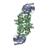
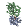
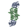
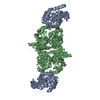
| Title | CRYSTAL STRUCTURE OF WILD-TYPE TRYPTOPHAN SYNTHASE COMPLEXED WITH N-[1H-INDOL-3-YL-ACETYL]ASPARTIC ACID | ||||||
 Components Components | (Tryptophan Synthase ...) x 2 | ||||||
 Keywords Keywords | LYASE / CARBON-OXYGEN LYASE / TRYPTOPHAN BIOSYNTHESIS / PYRIDOXAL PHOSPHATE | ||||||
| Function / homology |  Function and homology information Function and homology informationtryptophan synthase / tryptophan synthase activity / L-tryptophan biosynthetic process / identical protein binding / cytosol / cytoplasm Similarity search - Function | ||||||
| Biological species |  Salmonella typhimurium (bacteria) Salmonella typhimurium (bacteria) | ||||||
| Method |  X-RAY DIFFRACTION / SYNCHROTRON / MOLECULAR REPLACEMENT / Resolution: 1.7 Å X-RAY DIFFRACTION / SYNCHROTRON / MOLECULAR REPLACEMENT / Resolution: 1.7 Å | ||||||
 Authors Authors | Weyand, M. / Schlichting, I. / Marabotti, A. / Mozzarelli, A. | ||||||
 Citation Citation | Journal: J.Biol.Chem. / Year: 2002 Title: Crystal structures of a new class of allosteric effectors complexed to tryptophan synthase. Authors: Weyand, M. / Schlichting, I. / Marabotti, A. / Mozzarelli, A. | ||||||
| History |
|
- Structure visualization
| Structure viewer | Molecule: MolmilJmol/JSmol |
|---|
- Downloads & links
-Download
| PDBx/mmCIF format | 1k3u.cif.gz | 157 KB | Display | PDBx/mmCIF format |
|---|---|---|---|---|
| PDB format | pdb1k3u.ent.gz | 119.5 KB | Display | PDB format |
| PDBx/mmJSON format | 1k3u.json.gz | Tree view | PDBx/mmJSON format | |
| Others |  Other downloads Other downloads |
-Validation report
| Arichive directory | https://data.pdbj.org/pub/pdb/validation_reports/k3/1k3uftp://data.pdbj.org/pub/pdb/validation_reports/k3/1k3u | HTTPS FTP |
|---|
-Related structure data
| Related structure data |  1k7eC  1k7fC  1qopS C: citing same article ( S: Starting model for refinement |
|---|---|
| Similar structure data |


-Links
 PDBj
PDBj
- Assembly
| Deposited unit | 
| |||||||||
|---|---|---|---|---|---|---|---|---|---|---|
| 1 | 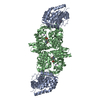
| |||||||||
| Unit cell |
| |||||||||
| Components on special symmetry positions |
|
-Components
-Tryptophan Synthase ... , 2 types, 2 molecules AB
| #1: Protein | Mass: 28698.797 Da / Num. of mol.: 1 Source method: isolated from a genetically manipulated source Source: (gene. exp.) Salmonella typhimurium (bacteria) / Gene: TRPA/TRPB / Plasmid: PSTB7 / Production host: |
|---|---|
| #2: Protein | Mass: 42787.688 Da / Num. of mol.: 1 Source method: isolated from a genetically manipulated source Source: (gene. exp.) Salmonella typhimurium (bacteria) / Gene: TRPA/TRPB / Plasmid: PSTB7 / Production host: |
-Non-polymers , 4 types, 715 molecules 


| #3: Chemical | ChemComp-IAD /  Mass: 290.271 Da / Num. of mol.: 1 / Source method: obtained synthetically / Formula: C14H14N2O5 Mass: 290.271 Da / Num. of mol.: 1 / Source method: obtained synthetically / Formula: C14H14N2O5 |
|---|---|
| #4: Chemical | ChemComp-NA /  Mass: 22.990 Da / Num. of mol.: 1 / Source method: obtained synthetically / Formula: Na Mass: 22.990 Da / Num. of mol.: 1 / Source method: obtained synthetically / Formula: Na |
| #5: Chemical | ChemComp-PLP /  Mass: 247.142 Da / Num. of mol.: 1 / Source method: obtained synthetically / Formula: C8H10NO6P Mass: 247.142 Da / Num. of mol.: 1 / Source method: obtained synthetically / Formula: C8H10NO6P |
| #6: Water | ChemComp-HOH / Mass: 18.015 Da / Num. of mol.: 712 / Source method: isolated from a natural source / Formula: H2O |
-Experimental details
-Experiment
| Experiment | Method: X-RAY DIFFRACTION / Number of used crystals: 1 |
|---|
- Sample preparation
| Crystal | Density Matthews: 2.55 Å3/Da / Density % sol: 51.76 % | ||||||||||||||||||||||||||||||||||||||||||||||||||||||||||||
|---|---|---|---|---|---|---|---|---|---|---|---|---|---|---|---|---|---|---|---|---|---|---|---|---|---|---|---|---|---|---|---|---|---|---|---|---|---|---|---|---|---|---|---|---|---|---|---|---|---|---|---|---|---|---|---|---|---|---|---|---|---|
| Crystal grow | Temperature: 293 K / Method: vapor diffusion, hanging drop / pH: 7.8 Details: PEG 8000, pH 7.8, VAPOR DIFFUSION, HANGING DROP, temperature 293K | ||||||||||||||||||||||||||||||||||||||||||||||||||||||||||||
| Crystal grow | *PLUS | ||||||||||||||||||||||||||||||||||||||||||||||||||||||||||||
| Components of the solutions | *PLUS
|
-Data collection
| Diffraction | Mean temperature: 100 K |
|---|---|
| Diffraction source | Source: SYNCHROTRON / Site: EMBL/DESY, HAMBURG  / Beamline: X11 / Wavelength: 0.842 / Beamline: X11 / Wavelength: 0.842 |
| Detector | Type: MAR scanner 345 mm plate / Detector: IMAGE PLATE / Date: Aug 10, 2000 / Details: Premirror, Bent mirror |
| Radiation | Monochromator: Triangular monochromator / Protocol: SINGLE WAVELENGTH / Monochromatic (M) / Laue (L): M / Scattering type: x-ray |
| Radiation wavelength | Wavelength: 0.842 Å / Relative weight: 1 |
| Reflection | Resolution: 1.7→31 Å / Num. obs: 77570 / % possible obs: 97.7 % / Observed criterion σ(F): -3 / Observed criterion σ(I): -3 / Redundancy: 3.05 % / Rsym value: 0.048 / Net I/σ(I): 14.4 |
| Reflection shell | Resolution: 1.7→1.8 Å / Mean I/σ(I) obs: 2.5 / Rsym value: 0.123 / % possible all: 94.2 |
| Reflection | *PLUS Lowest resolution: 31 Å / Num. measured all: 236589 / Rmerge(I) obs: 0.048 |
| Reflection shell | *PLUS % possible obs: 94.2 % / Rmerge(I) obs: 0.123 / Mean I/σ(I) obs: 2.5 |
- Processing
| Software |
| ||||||||||||||||||||||||||||||||||||||||||||||||||||||||||||||||||||||||||||||||||||||||||||||||||||||||
|---|---|---|---|---|---|---|---|---|---|---|---|---|---|---|---|---|---|---|---|---|---|---|---|---|---|---|---|---|---|---|---|---|---|---|---|---|---|---|---|---|---|---|---|---|---|---|---|---|---|---|---|---|---|---|---|---|---|---|---|---|---|---|---|---|---|---|---|---|---|---|---|---|---|---|---|---|---|---|---|---|---|---|---|---|---|---|---|---|---|---|---|---|---|---|---|---|---|---|---|---|---|---|---|---|---|
| Refinement | Method to determine structure: MOLECULAR REPLACEMENT Starting model: 1QOP Resolution: 1.7→20 Å / SU B: 1.91043 / SU ML: 0.06314 / Cross valid method: THROUGHOUT / σ(F): 0 / ESU R: 0.09026 / ESU R Free: 0.09174 / Stereochemistry target values: Engh & Huber Details: GLU 11 B exists in two conformations, each with 50% occupancy. With conformation A, HOH 709 exists. With conformation B, HOH 710 exists.
| ||||||||||||||||||||||||||||||||||||||||||||||||||||||||||||||||||||||||||||||||||||||||||||||||||||||||
| Solvent computation | Ion probe radii: 0.8 Å / Shrinkage radii: 0.8 Å / VDW probe radii: 1.4 Å | ||||||||||||||||||||||||||||||||||||||||||||||||||||||||||||||||||||||||||||||||||||||||||||||||||||||||
| Displacement parameters | Biso mean: 18.909 Å2
| ||||||||||||||||||||||||||||||||||||||||||||||||||||||||||||||||||||||||||||||||||||||||||||||||||||||||
| Refinement step | Cycle: LAST / Resolution: 1.7→20 Å
| ||||||||||||||||||||||||||||||||||||||||||||||||||||||||||||||||||||||||||||||||||||||||||||||||||||||||
| Refine LS restraints |
| ||||||||||||||||||||||||||||||||||||||||||||||||||||||||||||||||||||||||||||||||||||||||||||||||||||||||
| LS refinement shell | Resolution: 1.7→1.8 Å / % reflection obs: 94.2 % | ||||||||||||||||||||||||||||||||||||||||||||||||||||||||||||||||||||||||||||||||||||||||||||||||||||||||
| Refinement | *PLUS Lowest resolution: 20 Å / % reflection Rfree: 5 % / Rfactor obs: 0.1547 / Rfactor Rfree: 0.188 / Rfactor Rwork: 0.153 | ||||||||||||||||||||||||||||||||||||||||||||||||||||||||||||||||||||||||||||||||||||||||||||||||||||||||
| Solvent computation | *PLUS | ||||||||||||||||||||||||||||||||||||||||||||||||||||||||||||||||||||||||||||||||||||||||||||||||||||||||
| Displacement parameters | *PLUS | ||||||||||||||||||||||||||||||||||||||||||||||||||||||||||||||||||||||||||||||||||||||||||||||||||||||||
| Refine LS restraints | *PLUS
|
