 Movie
Movie Controller

[English] 日本語
 Yorodumi
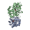
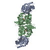
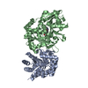
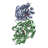
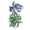
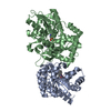
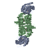
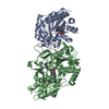
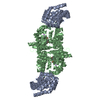
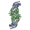
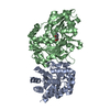
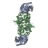
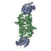
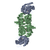
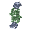
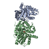
- PDB-1k7e: CRYSTAL STRUCTURE OF WILD-TYPE TRYPTOPHAN SYNTHASE COMPLEXED WITH... -
+ Open data

- Basic information
| Entry | Database: PDB / ID: 1k7e | ||||||
|---|---|---|---|---|---|---|---|
| Title | CRYSTAL STRUCTURE OF WILD-TYPE TRYPTOPHAN SYNTHASE COMPLEXED WITH N-[1H-INDOL-3-YL-ACETYL]GLYCINE ACID | ||||||
 Components Components | (TRYPTOPHAN SYNTHASE ...) x 2 | ||||||
 Keywords Keywords | LYASE / CARBON-OXYGEN LYASE / TRYPTOPHAN BIOSYNTHESIS / PYRIDOXAL PHOSPHATE | ||||||
| Function / homology |  Function and homology information Function and homology informationtryptophan synthase / tryptophan synthase activity / L-tryptophan biosynthetic process / identical protein binding / cytoplasm / cytosol Similarity search - Function | ||||||
| Biological species |  Salmonella typhimurium (bacteria) Salmonella typhimurium (bacteria) | ||||||
| Method |  X-RAY DIFFRACTION / SYNCHROTRON / MOLECULAR REPLACEMENT / Resolution: 2.3 Å X-RAY DIFFRACTION / SYNCHROTRON / MOLECULAR REPLACEMENT / Resolution: 2.3 Å | ||||||
 Authors Authors | Weyand, M. / Schlichting, I. / Marabotti, A. / Mozzarelli, A. | ||||||
 Citation Citation | Journal: J.Biol.Chem. / Year: 2002 Title: Crystal structures of a new class of allosteric effectors complexed to tryptophan synthase. Authors: Weyand, M. / Schlichting, I. / Marabotti, A. / Mozzarelli, A. | ||||||
| History |
|
- Structure visualization
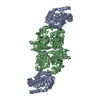
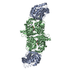
| Structure viewer | Molecule: MolmilJmol/JSmol |
|---|
- Downloads & links
-Download
| PDBx/mmCIF format | 1k7e.cif.gz | 145.2 KB | Display | PDBx/mmCIF format |
|---|---|---|---|---|
| PDB format | pdb1k7e.ent.gz | 109.3 KB | Display | PDB format |
| PDBx/mmJSON format | 1k7e.json.gz | Tree view | PDBx/mmJSON format | |
| Others |  Other downloads Other downloads |
-Validation report
| Arichive directory | https://data.pdbj.org/pub/pdb/validation_reports/k7/1k7eftp://data.pdbj.org/pub/pdb/validation_reports/k7/1k7e | HTTPS FTP |
|---|
-Related structure data
| Related structure data |  1k3uC  1k7fC  1qopS C: citing same article ( S: Starting model for refinement |
|---|---|
| Similar structure data |


-Links
 PDBj
PDBj
- Assembly
| Deposited unit | 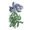
| ||||||||
|---|---|---|---|---|---|---|---|---|---|
| 1 | 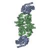
| ||||||||
| Unit cell |
|
-Components
-TRYPTOPHAN SYNTHASE ... , 2 types, 2 molecules AB
| #1: Protein | Mass: 28698.797 Da / Num. of mol.: 1 Source method: isolated from a genetically manipulated source Source: (gene. exp.) Salmonella typhimurium (bacteria) / Gene: TRPA/TRPB / Plasmid: PSTB7 / Production host: |
|---|---|
| #2: Protein | Mass: 42787.688 Da / Num. of mol.: 1 Source method: isolated from a genetically manipulated source Source: (gene. exp.) Salmonella typhimurium (bacteria) / Gene: TRPA/TRPB / Plasmid: PSTB7 / Production host: |
-Non-polymers , 4 types, 240 molecules 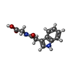


| #3: Chemical | ChemComp-IAG /  Mass: 232.235 Da / Num. of mol.: 1 / Source method: obtained synthetically / Formula: C12H12N2O3 Mass: 232.235 Da / Num. of mol.: 1 / Source method: obtained synthetically / Formula: C12H12N2O3 |
|---|---|
| #4: Chemical | ChemComp-NA /  Mass: 22.990 Da / Num. of mol.: 1 / Source method: obtained synthetically / Formula: Na Mass: 22.990 Da / Num. of mol.: 1 / Source method: obtained synthetically / Formula: Na |
| #5: Chemical | ChemComp-PLP /  Mass: 247.142 Da / Num. of mol.: 1 / Source method: obtained synthetically / Formula: C8H10NO6P Mass: 247.142 Da / Num. of mol.: 1 / Source method: obtained synthetically / Formula: C8H10NO6P |
| #6: Water | ChemComp-HOH / Mass: 18.015 Da / Num. of mol.: 237 / Source method: isolated from a natural source / Formula: H2O |
-Experimental details
-Experiment
| Experiment | Method: X-RAY DIFFRACTION / Number of used crystals: 1 |
|---|
- Sample preparation
| Crystal | Density Matthews: 2.48 Å3/Da / Density % sol: 50 % | ||||||||||||||||||||||||||||||||||||||||||||||||||||||||||||
|---|---|---|---|---|---|---|---|---|---|---|---|---|---|---|---|---|---|---|---|---|---|---|---|---|---|---|---|---|---|---|---|---|---|---|---|---|---|---|---|---|---|---|---|---|---|---|---|---|---|---|---|---|---|---|---|---|---|---|---|---|---|
| Crystal grow | pH: 7.8 / Details: PEG 8000, pH 7.80 | ||||||||||||||||||||||||||||||||||||||||||||||||||||||||||||
| Crystal grow | *PLUS pH: 7.8 / Method: vapor diffusion, hanging drop | ||||||||||||||||||||||||||||||||||||||||||||||||||||||||||||
| Components of the solutions | *PLUS
|
-Data collection
| Diffraction | Mean temperature: 100 K |
|---|---|
| Diffraction source | Source: SYNCHROTRON / Site: EMBL/DESY, HAMBURG  / Beamline: BW7B / Wavelength: 0.84 / Beamline: BW7B / Wavelength: 0.84 |
| Detector | Type: MAR scanner 345 mm plate / Detector: IMAGE PLATE / Date: Aug 10, 2000 / Details: PREMIRROR, BENT MIRROR |
| Radiation | Monochromator: TRIANGULAR MONOCHROMATOR / Protocol: SINGLE WAVELENGTH / Monochromatic (M) / Laue (L): M / Scattering type: x-ray |
| Radiation wavelength | Wavelength: 0.84 Å / Relative weight: 1 |
| Reflection | Resolution: 2.3→35 Å / % possible obs: 91 % / Observed criterion σ(I): -3 / Redundancy: 3.26 % / Rsym value: 0.058 / Net I/σ(I): 11.3 |
| Reflection shell | Resolution: 2.3→2.4 Å / Mean I/σ(I) obs: 2.2 / Rsym value: 0.183 / % possible all: 88.7 |
| Reflection | *PLUS Highest resolution: 2.3 Å / Lowest resolution: 35 Å / Num. obs: 29178 / Observed criterion σ(I): -3 / Num. measured all: 95120 / Rmerge(I) obs: 0.058 |
| Reflection shell | *PLUS Highest resolution: 2.3 Å / Lowest resolution: 2.4 Å / % possible obs: 88.7 % / Rmerge(I) obs: 0.183 / Mean I/σ(I) obs: 2.2 |
- Processing
| Software |
| ||||||||||||||||||||||||||||||||||||||||||||||||||||||||||||||||||||||||||||||||||||
|---|---|---|---|---|---|---|---|---|---|---|---|---|---|---|---|---|---|---|---|---|---|---|---|---|---|---|---|---|---|---|---|---|---|---|---|---|---|---|---|---|---|---|---|---|---|---|---|---|---|---|---|---|---|---|---|---|---|---|---|---|---|---|---|---|---|---|---|---|---|---|---|---|---|---|---|---|---|---|---|---|---|---|---|---|---|
| Refinement | Method to determine structure: MOLECULAR REPLACEMENT Starting model: 1QOP Resolution: 2.3→20 Å / Cross valid method: THROUGHOUT / σ(F): 0
| ||||||||||||||||||||||||||||||||||||||||||||||||||||||||||||||||||||||||||||||||||||
| Displacement parameters | Biso mean: 20.513 Å2
| ||||||||||||||||||||||||||||||||||||||||||||||||||||||||||||||||||||||||||||||||||||
| Refinement step | Cycle: LAST / Resolution: 2.3→20 Å
| ||||||||||||||||||||||||||||||||||||||||||||||||||||||||||||||||||||||||||||||||||||
| Refine LS restraints |
| ||||||||||||||||||||||||||||||||||||||||||||||||||||||||||||||||||||||||||||||||||||
| Refinement | *PLUS Highest resolution: 2.3 Å / % reflection Rfree: 5 % / Rfactor obs: 0.171 / Rfactor Rfree: 0.244 / Rfactor Rwork: 0.166 | ||||||||||||||||||||||||||||||||||||||||||||||||||||||||||||||||||||||||||||||||||||
| Solvent computation | *PLUS | ||||||||||||||||||||||||||||||||||||||||||||||||||||||||||||||||||||||||||||||||||||
| Displacement parameters | *PLUS | ||||||||||||||||||||||||||||||||||||||||||||||||||||||||||||||||||||||||||||||||||||
| Refine LS restraints | *PLUS
|
