 Movie
Movie Controller

[English] 日本語
 Yorodumi
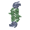
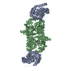
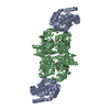
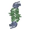
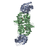
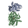
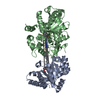
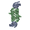
- PDB-1c29: CRYSTAL STRUCTURE OF THE COMPLEX OF BACTERIAL TRYPTOPHAN SYNTHASE... -
+ Open data

- Basic information
| Entry | Database: PDB / ID: 1c29 | ||||||
|---|---|---|---|---|---|---|---|
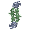
| Title | CRYSTAL STRUCTURE OF THE COMPLEX OF BACTERIAL TRYPTOPHAN SYNTHASE WITH THE TRANSITION STATE ANALOGUE INHIBITOR 4-(2-HYDROXYPHENYLTHIO)-1-BUTENYLPHOSPHONIC ACID | ||||||
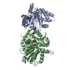
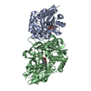
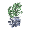
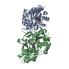
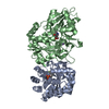
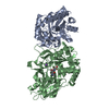
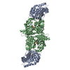
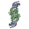
 Components Components | (TRYPTOPHAN SYNTHASE) x 2 | ||||||
 Keywords Keywords | LYASE/LYASE INHIBITOR / 8-FOLD ALPHA-BETA BARREL / ENZYME-INHIBITOR COMPLEX / LYASE-LYASE INHIBITOR COMPLEX | ||||||
| Function / homology |  Function and homology information Function and homology informationtryptophan synthase / tryptophan synthase activity / L-tryptophan biosynthetic process / identical protein binding / cytoplasm / cytosol Similarity search - Function | ||||||
| Biological species |  Salmonella typhimurium (bacteria) Salmonella typhimurium (bacteria) | ||||||
| Method |  X-RAY DIFFRACTION / Resolution: 2.3 Å X-RAY DIFFRACTION / Resolution: 2.3 Å | ||||||
 Authors Authors | Sachpatzidis, A. / Dealwis, C. / Lubetsky, J.B. / Liang, P.H. / Anderson, K.S. / Lolis, E. | ||||||
 Citation Citation | Journal: Biochemistry / Year: 1999 Title: Crystallographic studies of phosphonate-based alpha-reaction transition-state analogues complexed to tryptophan synthase. Authors: Sachpatzidis, A. / Dealwis, C. / Lubetsky, J.B. / Liang, P.H. / Anderson, K.S. / Lolis, E. #1: Journal: J.Biol.Chem. / Year: 1988Title: Three-Dimensional Structure of the Tryptophan Synthase Alpha 2 Beta 2 Multienzyme Complex from Salmonella typhimurium Authors: Hyde, C.C. / Ahmed, S.A. / Padlan, E.A. / Miles, E.W. / Davies, D.R. #2: Journal: Biochemistry / Year: 1998Title: Loop Closure and Intersubunit Communication in Tryptophan Synthase Authors: Schneider, T.R. / Gerhardt, E. / Lee, M. / Liang, P.H. / Anderson, K.S. / Schlichting, I. | ||||||
| History |
|
- Structure visualization
| Structure viewer | Molecule: MolmilJmol/JSmol |
|---|
- Downloads & links
-Download
| PDBx/mmCIF format | 1c29.cif.gz | 136.8 KB | Display | PDBx/mmCIF format |
|---|---|---|---|---|
| PDB format | pdb1c29.ent.gz | 105.7 KB | Display | PDB format |
| PDBx/mmJSON format | 1c29.json.gz | Tree view | PDBx/mmJSON format | |
| Others |  Other downloads Other downloads |
-Validation report
| Arichive directory | https://data.pdbj.org/pub/pdb/validation_reports/c2/1c29ftp://data.pdbj.org/pub/pdb/validation_reports/c2/1c29 | HTTPS FTP |
|---|
-Related structure data


-Links
 PDBj
PDBj
- Assembly
| Deposited unit | 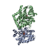
| ||||||||
|---|---|---|---|---|---|---|---|---|---|
| 1 | 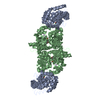
| ||||||||
| Unit cell |
|
-Components
-Protein , 2 types, 2 molecules AB
| #1: Protein | Mass: 28698.797 Da / Num. of mol.: 1 / Fragment: ALPHA CHAIN Source method: isolated from a genetically manipulated source Source: (gene. exp.) Salmonella typhimurium (bacteria) / Production host: |
|---|---|
| #2: Protein | Mass: 42988.996 Da / Num. of mol.: 1 / Fragment: BETA CHAIN Source method: isolated from a genetically manipulated source Source: (gene. exp.) Salmonella typhimurium (bacteria) / Production host: |
-Non-polymers , 4 types, 165 molecules 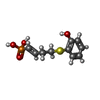


| #3: Chemical | ChemComp-HE1 /  Mass: 260.247 Da / Num. of mol.: 1 / Source method: obtained synthetically / Formula: C10H13O4PS Mass: 260.247 Da / Num. of mol.: 1 / Source method: obtained synthetically / Formula: C10H13O4PSDetails: INHIBITOR OF THE ALPHA-REACTION OF TRYPTOPHAN SYNTHASE NONCOVALENTLY BOUND AT THE ALPHA SUBUNIT ACTIVE SITE |
|---|---|
| #4: Chemical | ChemComp-NA /  Mass: 22.990 Da / Num. of mol.: 1 / Source method: obtained synthetically / Formula: Na Mass: 22.990 Da / Num. of mol.: 1 / Source method: obtained synthetically / Formula: NaDetails: METAL COFACTOR BOUND AT THE BETA SUBUNIT ACTIVE SITE |
| #5: Chemical | ChemComp-PLP /  Mass: 247.142 Da / Num. of mol.: 1 / Source method: obtained synthetically / Formula: C8H10NO6P Mass: 247.142 Da / Num. of mol.: 1 / Source method: obtained synthetically / Formula: C8H10NO6PDetails: NATURAL COFACTOR OF TRYPTOPHAN SYNTHASE BOUND AS A SCHIFF BASE WITH BETA LYS- 87 AT THE BETA SUBUNIT ACTIVE SITE |
| #6: Water | ChemComp-HOH / Mass: 18.015 Da / Num. of mol.: 162 / Source method: isolated from a natural source / Formula: H2O |
-Experimental details
-Experiment
| Experiment | Method: X-RAY DIFFRACTION / Number of used crystals: 1 |
|---|
- Sample preparation
| Crystal | Density Matthews: 2.51 Å3/Da / Density % sol: 51.07 % | |||||||||||||||||||||||||||||||||||
|---|---|---|---|---|---|---|---|---|---|---|---|---|---|---|---|---|---|---|---|---|---|---|---|---|---|---|---|---|---|---|---|---|---|---|---|---|
| Crystal grow | Temperature: 295 K / Method: vapor diffusion, hanging drop / pH: 7.8 Details: 12% PEG 4000, 0.75MM SPERMINE, 50MM SODIUM BICINE, 1MM EDTA, 5MM DTT, pH 7.8, VAPOR DIFFUSION, HANGING DROP, temperature 295K | |||||||||||||||||||||||||||||||||||
| Crystal grow | *PLUS Method: vapor diffusion / Details: pH was adjusted with NaOH | |||||||||||||||||||||||||||||||||||
| Components of the solutions | *PLUS
|
-Data collection
| Diffraction | Mean temperature: 140 K |
|---|---|
| Diffraction source | Source: ROTATING ANODE / Type: RIGAKU RU200 / Wavelength: 1.5418 |
| Detector | Type: RIGAKU RAXIS IIC / Detector: IMAGE PLATE / Date: May 27, 1997 |
| Radiation | Protocol: SINGLE WAVELENGTH / Monochromatic (M) / Laue (L): M / Scattering type: x-ray |
| Radiation wavelength | Wavelength: 1.5418 Å / Relative weight: 1 |
| Reflection | Resolution: 2.3→42.2 Å / Num. all: 30087 / Num. obs: 29830 / % possible obs: 92.6 % / Redundancy: 2.1 % / Biso Wilson estimate: 11.63 Å2 / Rmerge(I) obs: 0.076 / Net I/σ(I): 12.9 |
| Reflection shell | Resolution: 2.3→2.4 Å / Redundancy: 2.1 % / Rmerge(I) obs: 0.174 / % possible all: 93 |
| Reflection | *PLUS Num. measured all: 245222 |
| Reflection shell | *PLUS % possible obs: 93 % |
- Processing
| Software |
| ||||||||||||||||||||||||||||||||||||||||||||||||||||||||||||
|---|---|---|---|---|---|---|---|---|---|---|---|---|---|---|---|---|---|---|---|---|---|---|---|---|---|---|---|---|---|---|---|---|---|---|---|---|---|---|---|---|---|---|---|---|---|---|---|---|---|---|---|---|---|---|---|---|---|---|---|---|---|
| Refinement | Resolution: 2.3→30 Å / σ(F): 2 / Stereochemistry target values: ENGH & HUBER Details: RESTRAINED LEAST SQUARES REFINEMENT. CONJUGATE GRADIENT MINIMIZATION AND SIMULATED ANNEALING PROTOCOLS IMPLEMENTED IN XPLOR.
| ||||||||||||||||||||||||||||||||||||||||||||||||||||||||||||
| Refine analyze | Luzzati sigma a obs: 0.25 Å | ||||||||||||||||||||||||||||||||||||||||||||||||||||||||||||
| Refinement step | Cycle: LAST / Resolution: 2.3→30 Å
| ||||||||||||||||||||||||||||||||||||||||||||||||||||||||||||
| Refine LS restraints |
| ||||||||||||||||||||||||||||||||||||||||||||||||||||||||||||
| Software | *PLUS Name: X-PLOR / Version: 3.851 / Classification: refinement | ||||||||||||||||||||||||||||||||||||||||||||||||||||||||||||
| Refinement | *PLUS | ||||||||||||||||||||||||||||||||||||||||||||||||||||||||||||
| Solvent computation | *PLUS | ||||||||||||||||||||||||||||||||||||||||||||||||||||||||||||
| Displacement parameters | *PLUS | ||||||||||||||||||||||||||||||||||||||||||||||||||||||||||||
| Refine LS restraints | *PLUS
|
