Mass: 18.015 Da / Num. of mol.: 483 / Source method: isolated from a natural source / Formula: H2O
-
Details
Has protein modification
N
-
Experimental details
-
Experiment
Experiment
Method: X-RAY DIFFRACTION / Number of used crystals: 1
-
Sample preparation
Crystal
Density Matthews: 2.54 Å3/Da / Density % sol: 51.63 %
Crystal grow
Temperature: 293 K / Method: vapor diffusion, hanging drop / pH: 9 Details: Protein: 50 mM Bicine pH 7.8, 10 mM Na-EDTA, 1 mM DTE, 20 uM PLP. Reservoir: 50 mM Bicine pH 7.8, 5 mM DTE, 5 mM Na-EDTA, 0.1 M PLP, 2 mM Spermine, 2 mM NaN3, 8-12% PEG 8000. Crystal soaked ...Details: Protein: 50 mM Bicine pH 7.8, 10 mM Na-EDTA, 1 mM DTE, 20 uM PLP. Reservoir: 50 mM Bicine pH 7.8, 5 mM DTE, 5 mM Na-EDTA, 0.1 M PLP, 2 mM Spermine, 2 mM NaN3, 8-12% PEG 8000. Crystal soaked in 15% PEG 8000, 20% Glycerol, 23 mM IGP, pH controlled at pH 9.0 before flash-cooling, VAPOR DIFFUSION, HANGING DROP, temperature 293K
Protocol: SINGLE WAVELENGTH / Monochromatic (M) / Laue (L): M / Scattering type: x-ray
Radiation wavelength
Wavelength: 0.93 Å / Relative weight: 1
Reflection
Resolution: 1.7→55 Å / Num. all: 77139 / Num. obs: 77139 / % possible obs: 97.3 % / Observed criterion σ(I): -3 / Redundancy: 2.4 % / Biso Wilson estimate: 19.8 Å2 / Rmerge(I) obs: 0.055 / Net I/σ(I): 10.01
Reflection shell
Resolution: 1.7→1.8 Å / Rmerge(I) obs: 0.313 / Mean I/σ(I) obs: 2.46 / Num. unique all: 12104 / % possible all: 97.1
-
Processing
Software
Name
Version
Classification
REFMAC
5.2.0005
refinement
ADSC
Quantum
datacollection
XDS
datareduction
XSCALE
datascaling
CNS
phasing
Refinement
Method to determine structure: FOURIER SYNTHESIS / Resolution: 1.7→43.73 Å / Cor.coef. Fo:Fc: 0.963 / Cor.coef. Fo:Fc free: 0.951 / SU B: 2.066 / SU ML: 0.068 / Cross valid method: THROUGHOUT / σ(F): 0 / ESU R: 0.097 / ESU R Free: 0.095 / Stereochemistry target values: MAXIMUM LIKELIHOOD Details: HYDROGENS HAVE BEEN ADDED IN THE RIDING POSITIONS. Program CNS has also been used in refinement
Rfactor
Num. reflection
% reflection
Selection details
Rfree
0.20044
3922
5.1 %
RANDOM
Rwork
0.17194
-
-
-
all
0.17338
73246
-
-
obs
0.17338
73246
100 %
-
Solvent computation
Ion probe radii: 0.8 Å / Shrinkage radii: 0.8 Å / VDW probe radii: 1.2 Å / Solvent model: MASK
Displacement parameters
Biso mean: 20.991 Å2
Baniso -1
Baniso -2
Baniso -3
1-
-0.8 Å2
0 Å2
-0.41 Å2
2-
-
2.19 Å2
0 Å2
3-
-
-
-1.46 Å2
Refinement step
Cycle: LAST / Resolution: 1.7→43.73 Å
Protein
Nucleic acid
Ligand
Solvent
Total
Num. atoms
4895
0
35
483
5413
Refine LS restraints
Refine-ID
Type
Dev ideal
Dev ideal target
Number
X-RAY DIFFRACTION
r_bond_refined_d
0.011
0.022
5082
X-RAY DIFFRACTION
r_bond_other_d
0.001
0.02
4660
X-RAY DIFFRACTION
r_angle_refined_deg
1.426
1.979
6888
X-RAY DIFFRACTION
r_angle_other_deg
0.924
3
10841
X-RAY DIFFRACTION
r_dihedral_angle_1_deg
5.407
5
650
X-RAY DIFFRACTION
r_dihedral_angle_2_deg
35.48
24.11
219
X-RAY DIFFRACTION
r_dihedral_angle_3_deg
13.281
15
836
X-RAY DIFFRACTION
r_dihedral_angle_4_deg
19.246
15
32
X-RAY DIFFRACTION
r_chiral_restr
0.075
0.2
762
X-RAY DIFFRACTION
r_gen_planes_refined
0.005
0.02
5720
X-RAY DIFFRACTION
r_gen_planes_other
0.001
0.02
993
X-RAY DIFFRACTION
r_nbd_refined
0.219
0.2
1028
X-RAY DIFFRACTION
r_nbd_other
0.185
0.2
4620
X-RAY DIFFRACTION
r_nbtor_refined
0.177
0.2
2476
X-RAY DIFFRACTION
r_nbtor_other
0.084
0.2
2725
X-RAY DIFFRACTION
r_xyhbond_nbd_refined
0.131
0.2
378
X-RAY DIFFRACTION
r_metal_ion_refined
0.098
0.2
3
X-RAY DIFFRACTION
r_symmetry_vdw_refined
0.135
0.2
7
X-RAY DIFFRACTION
r_symmetry_vdw_other
0.227
0.2
58
X-RAY DIFFRACTION
r_symmetry_hbond_refined
0.115
0.2
11
X-RAY DIFFRACTION
r_mcbond_it
0.714
1.5
3236
X-RAY DIFFRACTION
r_mcbond_other
0.18
1.5
1332
X-RAY DIFFRACTION
r_mcangle_it
1.308
2
5164
X-RAY DIFFRACTION
r_scbond_it
2.09
3
1877
X-RAY DIFFRACTION
r_scangle_it
3.452
4.5
1724
LS refinement shell
Resolution: 1.7→1.744 Å / Total num. of bins used: 20 /
Rfactor
Num. reflection
Rfree
0.243
296
Rwork
0.239
5384
+
About Yorodumi
-
News
-
Feb 9, 2022. New format data for meta-information of EMDB entries
New format data for meta-information of EMDB entries
Version 3 of the EMDB header file is now the official format.
The previous official version 1.9 will be removed from the archive.
In the structure databanks used in Yorodumi, some data are registered as the other names, "COVID-19 virus" and "2019-nCoV". Here are the details of the virus and the list of structure data.
Jan 31, 2019. EMDB accession codes are about to change! (news from PDBe EMDB page)
EMDB accession codes are about to change! (news from PDBe EMDB page)
The allocation of 4 digits for EMDB accession codes will soon come to an end. Whilst these codes will remain in use, new EMDB accession codes will include an additional digit and will expand incrementally as the available range of codes is exhausted. The current 4-digit format prefixed with “EMD-” (i.e. EMD-XXXX) will advance to a 5-digit format (i.e. EMD-XXXXX), and so on. It is currently estimated that the 4-digit codes will be depleted around Spring 2019, at which point the 5-digit format will come into force.
The EM Navigator/Yorodumi systems omit the EMD- prefix.
Related info.:Q: What is EMD? / ID/Accession-code notation in Yorodumi/EM Navigator
Yorodumi is a browser for structure data from EMDB, PDB, SASBDB, etc.
This page is also the successor to EM Navigator detail page, and also detail information page/front-end page for Omokage search.
The word "yorodu" (or yorozu) is an old Japanese word meaning "ten thousand". "mi" (miru) is to see.
Related info.:EMDB / PDB / SASBDB / Comparison of 3 databanks / Yorodumi Search / Aug 31, 2016. New EM Navigator & Yorodumi / Yorodumi Papers / Jmol/JSmol / Function and homology information / Changes in new EM Navigator and Yorodumi
 Movie
Movie Controller

 Yorodumi
 Open data
 Basic information
 Components
 Keywords
 Function and homology information
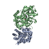
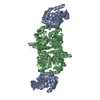
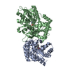
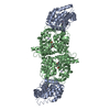
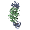
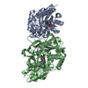
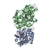
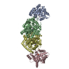
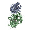
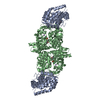
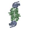
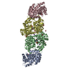
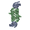
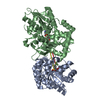
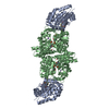
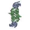
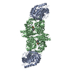
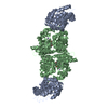
 Salmonella typhimurium (bacteria)
 X-RAY DIFFRACTION /
  Authors
 Citation
 Structure visualization
 Downloads & links
 Other downloads
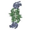


 PDBj
PDBj
 Assembly
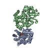


 Mass: 287.206 Da / Num. of mol.: 1 / Source method: obtained synthetically / Formula: C11H14NO6P
 Mass: 22.990 Da / Num. of mol.: 1 / Source method: obtained synthetically / Formula: Na
 Mass: 247.142 Da / Num. of mol.: 1 / Source method: obtained synthetically / Formula: C8H10NO6P
 Sample preparation
 / Beamline: ID14-3 / Wavelength: 0.93 Å
 Processing