 Movie
Movie Controller

[English] 日本語
 Yorodumi
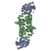
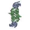
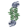
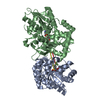
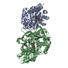
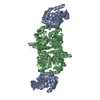
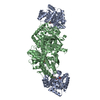
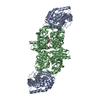
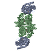
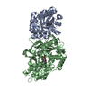
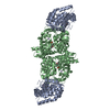
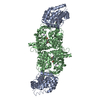
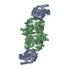
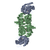
- PDB-1ttq: TRYPTOPHAN SYNTHASE (E.C.4.2.1.20) IN THE PRESENCE OF POTASSIUM A... -
+ Open data

- Basic information
| Entry | Database: PDB / ID: 1ttq | ||||||
|---|---|---|---|---|---|---|---|
| Title | TRYPTOPHAN SYNTHASE (E.C.4.2.1.20) IN THE PRESENCE OF POTASSIUM AT ROOM TEMPERATURE | ||||||
 Components Components | (TRYPTOPHAN SYNTHASE) x 2 | ||||||
 Keywords Keywords | CARBON-OXYGEN LYASE | ||||||
| Function / homology |  Function and homology information Function and homology informationtryptophan synthase / tryptophan synthase activity / L-tryptophan biosynthetic process / identical protein binding / cytosol / cytoplasm Similarity search - Function | ||||||
| Biological species |  Salmonella typhimurium (bacteria) Salmonella typhimurium (bacteria) | ||||||
| Method |  X-RAY DIFFRACTION / Resolution: 2 Å X-RAY DIFFRACTION / Resolution: 2 Å | ||||||
 Authors Authors | Rhee, S. / Parris, K. / Ahmed, S. / Miles, E.W. / Davies, D.R. | ||||||
 Citation Citation | Journal: Biochemistry / Year: 1996 Title: Exchange of K+ or Cs+ for Na+ induces local and long-range changes in the three-dimensional structure of the tryptophan synthase alpha2beta2 complex. Authors: Rhee, S. / Parris, K.D. / Ahmed, S.A. / Miles, E.W. / Davies, D.R. #1: Journal: Bio/Technology / Year: 1990Title: The Tryptophan Synthase Multienzyme Complex: Exploring Structure-Function Relationships with X-Ray Crystallography and Mutagenesis Authors: Hyde, C.C. / Miles, E.W. #2: Journal: J.Biol.Chem. / Year: 1988Title: Three-Dimensional Structure of the Tryptophan Synthase Alpha2Beta2 Multienzyme Complex from Salmonella Typhimurium Authors: Hyde, C.C. / Ahmed, S.A. / Padlan, E.A. / Miles, E.W. / Davies, D.R. #3: Journal: J.Biol.Chem. / Year: 1985Title: Crystallization and Preliminary X-Ray Crystallographic Data of the Tryptophan Synthase Alpha2Beta2 Complex from Salmonella Typhimurium Authors: Ahmed, S.A. / Miles, E.W. / Davies, D.R. | ||||||
| History |
|
- Structure visualization
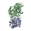
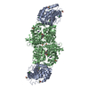
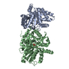
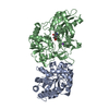
| Structure viewer | Molecule: MolmilJmol/JSmol |
|---|
- Downloads & links
-Download
| PDBx/mmCIF format | 1ttq.cif.gz | 135 KB | Display | PDBx/mmCIF format |
|---|---|---|---|---|
| PDB format | pdb1ttq.ent.gz | 104.8 KB | Display | PDB format |
| PDBx/mmJSON format | 1ttq.json.gz | Tree view | PDBx/mmJSON format | |
| Others |  Other downloads Other downloads |
-Validation report
| Arichive directory | https://data.pdbj.org/pub/pdb/validation_reports/tt/1ttqftp://data.pdbj.org/pub/pdb/validation_reports/tt/1ttq | HTTPS FTP |
|---|
-Related structure data


-Links
 PDBj
PDBj
- Assembly
| Deposited unit | 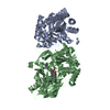
| ||||||||
|---|---|---|---|---|---|---|---|---|---|
| 1 | 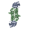
| ||||||||
| Unit cell |
| ||||||||
| Atom site foot note | 1: CIS PROLINE - PRO A 28 2: SER A 55 - ASP A 56 OMEGA = 148.59 PEPTIDE BOND DEVIATES SIGNIFICANTLY FROM TRANS CONFORMATION 3: GLY A 61 - PRO A 62 OMEGA = 216.22 PEPTIDE BOND DEVIATES SIGNIFICANTLY FROM TRANS CONFORMATION 4: SER A 247 - PRO A 248 OMEGA = 216.00 PEPTIDE BOND DEVIATES SIGNIFICANTLY FROM TRANS CONFORMATION 5: CIS PROLINE - PRO B 56 / 6: CIS PROLINE - PRO B 196 |
-Components
| #1: Protein | Mass: 28698.797 Da / Num. of mol.: 1 Source method: isolated from a genetically manipulated source Details: STRUCTURE IN THE PRESENCE OF POTASSIUM / Source: (gene. exp.) Salmonella typhimurium (bacteria) / Strain: TB2211 / Gene: TRPA/TRPB/TRPC / Plasmid: PSTH8 / Gene (production host): TRPA/TRPB/TRPC / Production host: |
|---|---|
| #2: Protein | Mass: 42988.996 Da / Num. of mol.: 1 Source method: isolated from a genetically manipulated source Details: STRUCTURE IN THE PRESENCE OF POTASSIUM / Source: (gene. exp.) Salmonella typhimurium (bacteria) / Strain: TB2211 / Gene: TRPA/TRPB/TRPC / Plasmid: PSTH8 / Gene (production host): TRPA/TRPB/TRPC / Production host: References: UniProt: P00933, UniProt: P0A2K1*PLUS, tryptophan synthase |
| #3: Chemical | ChemComp-K /   Mass: 39.098 Da / Num. of mol.: 1 / Source method: obtained synthetically / Formula: K Mass: 39.098 Da / Num. of mol.: 1 / Source method: obtained synthetically / Formula: K |
| #4: Chemical | ChemComp-PLP /   Mass: 247.142 Da / Num. of mol.: 1 / Source method: obtained synthetically / Formula: C8H10NO6P Mass: 247.142 Da / Num. of mol.: 1 / Source method: obtained synthetically / Formula: C8H10NO6P |
| #5: Water | ChemComp-HOH /  Mass: 18.015 Da / Num. of mol.: 75 / Source method: isolated from a natural source / Formula: H2O Mass: 18.015 Da / Num. of mol.: 75 / Source method: isolated from a natural source / Formula: H2O |
-Experimental details
-Experiment
| Experiment | Method: X-RAY DIFFRACTION |
|---|
- Sample preparation
| Crystal | Density Matthews: 2.67 Å3/Da / Density % sol: 53.88 % | |||||||||||||||||||||||||
|---|---|---|---|---|---|---|---|---|---|---|---|---|---|---|---|---|---|---|---|---|---|---|---|---|---|---|
| Crystal | *PLUS Density % sol: 48 % | |||||||||||||||||||||||||
| Crystal grow | *PLUS Temperature: 20 ℃ / pH: 7.8 / Method: unknown / Details: pH is adjusted to 7.8 with NaOH | |||||||||||||||||||||||||
| Components of the solutions | *PLUS
|
-Data collection
| Diffraction | Mean temperature: 293 K |
|---|---|
| Diffraction source | Wavelength: 1.5418 |
| Detector | Type: RIGAKU RAXIS IIC / Detector: IMAGE PLATE / Date: Nov 27, 1994 |
| Radiation | Monochromatic (M) / Laue (L): M / Scattering type: x-ray |
| Radiation wavelength | Wavelength: 1.5418 Å / Relative weight: 1 |
| Reflection | Resolution: 2→40 Å / Num. obs: 39626 / % possible obs: 77.8 % / Observed criterion σ(I): 0 / Redundancy: 2.5 % / Rmerge(I) obs: 0.079 |
| Reflection | *PLUS Num. measured all: 322940 / Rmerge(I) obs: 0.079 |
| Reflection shell | *PLUS Highest resolution: 2 Å / Lowest resolution: 2.07 Å / % possible obs: 42.4 % |
- Processing
| Software |
| ||||||||||||||||||||||||||||||||||||||||||||||||||||||||||||
|---|---|---|---|---|---|---|---|---|---|---|---|---|---|---|---|---|---|---|---|---|---|---|---|---|---|---|---|---|---|---|---|---|---|---|---|---|---|---|---|---|---|---|---|---|---|---|---|---|---|---|---|---|---|---|---|---|---|---|---|---|---|
| Refinement | Resolution: 2→8 Å / σ(F): 2
| ||||||||||||||||||||||||||||||||||||||||||||||||||||||||||||
| Refine analyze | Luzzati coordinate error obs: 0.3 Å | ||||||||||||||||||||||||||||||||||||||||||||||||||||||||||||
| Refinement step | Cycle: LAST / Resolution: 2→8 Å
| ||||||||||||||||||||||||||||||||||||||||||||||||||||||||||||
| Refine LS restraints |
| ||||||||||||||||||||||||||||||||||||||||||||||||||||||||||||
| Software | *PLUS Name: X-PLOR / Classification: refinement | ||||||||||||||||||||||||||||||||||||||||||||||||||||||||||||
| Refinement | *PLUS Rfactor obs: 0.204 / Rfactor Rfree: 0.289 | ||||||||||||||||||||||||||||||||||||||||||||||||||||||||||||
| Solvent computation | *PLUS | ||||||||||||||||||||||||||||||||||||||||||||||||||||||||||||
| Displacement parameters | *PLUS | ||||||||||||||||||||||||||||||||||||||||||||||||||||||||||||
| Refine LS restraints | *PLUS
|
