 Movie
Movie Controller


+ Open data

- Basic information
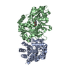
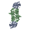
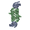
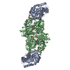
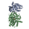
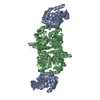
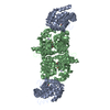
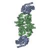
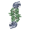
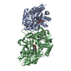
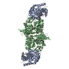
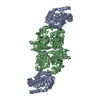
| Entry | Database: PDB / ID: 2wsy | ||||||
|---|---|---|---|---|---|---|---|
| Title | CRYSTAL STRUCTURE OF WILD-TYPE TRYPTOPHAN SYNTHASE | ||||||
 Components Components | (TRYPTOPHAN SYNTHASE) x 2 | ||||||
 Keywords Keywords | LYASE / CARBON-OXYGEN LYASE / TRYPTOPHAN BIOSYNTHESIS / PYRIDOXAL PHOSPHATE | ||||||
| Function / homology |  Function and homology information Function and homology informationtryptophan synthase / tryptophan synthase activity / L-tryptophan biosynthetic process / identical protein binding / cytosol / cytoplasm Similarity search - Function | ||||||
| Biological species |  Salmonella typhimurium (bacteria) Salmonella typhimurium (bacteria) | ||||||
| Method |  X-RAY DIFFRACTION / SYNCHROTRON / ISOMORPHOUS WITH PDB ENTRY 1WSY. / Resolution: 3.05 Å X-RAY DIFFRACTION / SYNCHROTRON / ISOMORPHOUS WITH PDB ENTRY 1WSY. / Resolution: 3.05 Å | ||||||
 Authors Authors | Schneider, T.R. / Gerhardt, E. / Lee, M. / Liang, P.-H. / Anderson, K.S. / Schlichting, I. | ||||||
 Citation Citation | Journal: Biochemistry / Year: 1998 Title: Loop closure and intersubunit communication in tryptophan synthase. Authors: Schneider, T.R. / Gerhardt, E. / Lee, M. / Liang, P.H. / Anderson, K.S. / Schlichting, I. | ||||||
| History |
|
- Structure visualization
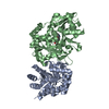
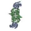
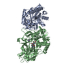
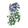
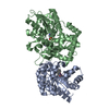
| Structure viewer | Molecule: MolmilJmol/JSmol |
|---|
- Downloads & links
-Download
| PDBx/mmCIF format | 2wsy.cif.gz | 121.5 KB | Display | PDBx/mmCIF format |
|---|---|---|---|---|
| PDB format | pdb2wsy.ent.gz | 96.4 KB | Display | PDB format |
| PDBx/mmJSON format | 2wsy.json.gz | Tree view | PDBx/mmJSON format | |
| Others |  Other downloads Other downloads |
-Validation report
| Arichive directory | https://data.pdbj.org/pub/pdb/validation_reports/ws/2wsyftp://data.pdbj.org/pub/pdb/validation_reports/ws/2wsy | HTTPS FTP |
|---|
-Related structure data
| Related structure data | 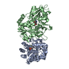 1a50C 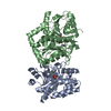 1a5sC  1wsy S: Starting model for refinement C: citing same article ( |
|---|---|
| Similar structure data |


-Links
 PDBj
PDBj
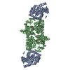
- Assembly
| Deposited unit | 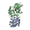
| ||||||||
|---|---|---|---|---|---|---|---|---|---|
| 1 | 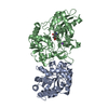
| ||||||||
| Unit cell |
|
-Components
| #1: Protein | Mass: 28698.797 Da / Num. of mol.: 1 / Source method: isolated from a natural source / Source: (natural) Salmonella typhimurium (bacteria) / Cell line: CB149 / Plasmid: PSTB7 / References: UniProt: P00929, tryptophan synthase |
|---|---|
| #2: Protein | Mass: 42857.805 Da / Num. of mol.: 1 / Source method: isolated from a natural source / Source: (natural) Salmonella typhimurium (bacteria) / Cell line: CB149 / Plasmid: PSTB7 / References: UniProt: P0A2K1, tryptophan synthase |
| #3: Chemical | ChemComp-NA /   Mass: 22.990 Da / Num. of mol.: 1 / Source method: obtained synthetically / Formula: Na Mass: 22.990 Da / Num. of mol.: 1 / Source method: obtained synthetically / Formula: Na |
| #4: Chemical | ChemComp-PLP /   Mass: 247.142 Da / Num. of mol.: 1 / Source method: obtained synthetically / Formula: C8H10NO6P Mass: 247.142 Da / Num. of mol.: 1 / Source method: obtained synthetically / Formula: C8H10NO6P |
-Experimental details
-Experiment
| Experiment | Method: X-RAY DIFFRACTION / Number of used crystals: 1 |
|---|
- Sample preparation
| Crystal | Density Matthews: 2.67 Å3/Da / Density % sol: 48 % | |||||||||||||||||||||||||||||||||||
|---|---|---|---|---|---|---|---|---|---|---|---|---|---|---|---|---|---|---|---|---|---|---|---|---|---|---|---|---|---|---|---|---|---|---|---|---|
| Crystal grow | pH: 7.8 / Details: pH 7.8 | |||||||||||||||||||||||||||||||||||
| Crystal grow | *PLUS Method: vapor diffusion, hanging dropDetails: drop consists of equal volume of enzyme and reservoir solutions | |||||||||||||||||||||||||||||||||||
| Components of the solutions | *PLUS
|
-Data collection
| Diffraction | Mean temperature: 277 K |
|---|---|
| Diffraction source | Source: SYNCHROTRON / Site: EMBL/DESY, HAMBURG  / Beamline: BW7B / Wavelength: 0.86 / Beamline: BW7B / Wavelength: 0.86 |
| Detector | Type: MARRESEARCH / Detector: IMAGE PLATE / Date: Nov 1, 1995 / Details: SYNCHROTRON |
| Radiation | Monochromator: SYNCHROTRON / Monochromatic (M) / Laue (L): M / Scattering type: x-ray |
| Radiation wavelength | Wavelength: 0.86 Å / Relative weight: 1 |
| Reflection | Resolution: 3.05→10 Å / Num. obs: 13162 / % possible obs: 90.9 % / Observed criterion σ(I): -3 / Redundancy: 2.1 % / Rmerge(I) obs: 0.034 / Net I/σ(I): 20.2 |
| Reflection shell | Highest resolution: 3.05 Å / Redundancy: 2.4 % / Rmerge(I) obs: 0.21 / Mean I/σ(I) obs: 4.2 / % possible all: 89.8 |
| Reflection | *PLUS Num. measured all: 28423 |
| Reflection shell | *PLUS % possible obs: 89.8 % |
- Processing
| Software |
| ||||||||||||||||||||||||||||||||||||||||||||||||||||||||||||
|---|---|---|---|---|---|---|---|---|---|---|---|---|---|---|---|---|---|---|---|---|---|---|---|---|---|---|---|---|---|---|---|---|---|---|---|---|---|---|---|---|---|---|---|---|---|---|---|---|---|---|---|---|---|---|---|---|---|---|---|---|---|
| Refinement | Method to determine structure: ISOMORPHOUS WITH PDB ENTRY 1WSY. Starting model: 1WSY 1wsy Resolution: 3.05→10 Å / Data cutoff high absF: 100000 / Data cutoff low absF: 0.001 / Isotropic thermal model: TIGHTLY RESTRAINED, SEE P / Cross valid method: THROUGHOUT / σ(F): 2
| ||||||||||||||||||||||||||||||||||||||||||||||||||||||||||||
| Displacement parameters | Biso mean: 20.4 Å2 | ||||||||||||||||||||||||||||||||||||||||||||||||||||||||||||
| Refine analyze | Luzzati d res low obs: 10 Å / Luzzati sigma a obs: 0.37 Å | ||||||||||||||||||||||||||||||||||||||||||||||||||||||||||||
| Refinement step | Cycle: LAST / Resolution: 3.05→10 Å
| ||||||||||||||||||||||||||||||||||||||||||||||||||||||||||||
| Refine LS restraints |
| ||||||||||||||||||||||||||||||||||||||||||||||||||||||||||||
| LS refinement shell | Resolution: 3.05→3.1 Å / Total num. of bins used: 20
| ||||||||||||||||||||||||||||||||||||||||||||||||||||||||||||
| Xplor file |
| ||||||||||||||||||||||||||||||||||||||||||||||||||||||||||||
| Software | *PLUS Name: X-PLOR / Version: 3.851 / Classification: refinement | ||||||||||||||||||||||||||||||||||||||||||||||||||||||||||||
| Refinement | *PLUS | ||||||||||||||||||||||||||||||||||||||||||||||||||||||||||||
| Solvent computation | *PLUS | ||||||||||||||||||||||||||||||||||||||||||||||||||||||||||||
| Displacement parameters | *PLUS | ||||||||||||||||||||||||||||||||||||||||||||||||||||||||||||
| LS refinement shell | *PLUS Rfactor obs: 0.3005 |
