 Movie
Movie Controller


+ Open data

- Basic information
| Entry | Database: PDB / ID: 2gr9 | ||||||
|---|---|---|---|---|---|---|---|
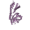
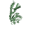
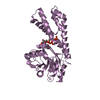
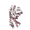
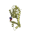
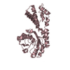
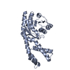
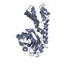
| Title | Crystal structure of P5CR complexed with NADH | ||||||
 Components Components | Pyrroline-5-carboxylate reductase 1 | ||||||
 Keywords Keywords | OXIDOREDUCTASE / crystal strucutre / Human Pyrroline-5-carboxylate Reductase / nadh | ||||||
| Function / homology |  Function and homology information Function and homology informationpyrroline-5-carboxylate reductase complex / pyrroline-5-carboxylate reductase / pyrroline-5-carboxylate reductase activity / Glutamate and glutamine metabolism / L-proline biosynthetic process / negative regulation of oxidative stress-induced neuron intrinsic apoptotic signaling pathway / regulation of mitochondrial membrane potential / cellular response to oxidative stress / mitochondrial matrix / mitochondrion / identical protein binding Similarity search - Function | ||||||
| Biological species |  Homo sapiens (human) Homo sapiens (human) | ||||||
| Method |  X-RAY DIFFRACTION / MOLECULAR REPLACEMENT / Resolution: 3.1 Å X-RAY DIFFRACTION / MOLECULAR REPLACEMENT / Resolution: 3.1 Å | ||||||
 Authors Authors | Meng, Z. / Lou, Z. / Liu, Z. / Rao, Z. | ||||||
 Citation Citation | Journal: J.Mol.Biol. / Year: 2006 Title: Crystal structure of human pyrroline-5-carboxylate reductase Authors: Meng, Z. / Lou, Z. / Liu, Z. / Li, M. / Zhao, X. / Bartlam, M. / Rao, Z. | ||||||
| History |
|
- Structure visualization
| Structure viewer | Molecule: MolmilJmol/JSmol |
|---|
- Downloads & links
-Download
| PDBx/mmCIF format | 2gr9.cif.gz | 287.4 KB | Display | PDBx/mmCIF format |
|---|---|---|---|---|
| PDB format | pdb2gr9.ent.gz | 229.5 KB | Display | PDB format |
| PDBx/mmJSON format | 2gr9.json.gz | Tree view | PDBx/mmJSON format | |
| Others |  Other downloads Other downloads |
-Validation report
| Arichive directory | https://data.pdbj.org/pub/pdb/validation_reports/gr/2gr9ftp://data.pdbj.org/pub/pdb/validation_reports/gr/2gr9 | HTTPS FTP |
|---|
-Related structure data
| Related structure data |  2gerSC 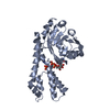 2graC C: citing same article ( S: Starting model for refinement |
|---|---|
| Similar structure data |


-Links
 PDBj
PDBj


- Assembly
| Deposited unit | 
| ||||||||
|---|---|---|---|---|---|---|---|---|---|
| 1 | 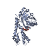
| ||||||||
| 2 | 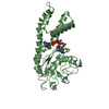
| ||||||||
| 3 | 
| ||||||||
| 4 | 
| ||||||||
| 5 | 
| ||||||||
| 6 | 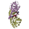
| ||||||||
| 7 | 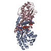
| ||||||||
| 8 | 
| ||||||||
| Unit cell |
|
-Components
| #1: Protein | Mass: 29147.648 Da / Num. of mol.: 5 Source method: isolated from a genetically manipulated source Source: (gene. exp.) Homo sapiens (human) / Production host:  References: UniProt: P32322, pyrroline-5-carboxylate reductase #2: Chemical | ChemComp-NAI /   Mass: 665.441 Da / Num. of mol.: 5 / Source method: obtained synthetically / Formula: C21H29N7O14P2 Mass: 665.441 Da / Num. of mol.: 5 / Source method: obtained synthetically / Formula: C21H29N7O14P2#3: Chemical | ChemComp-GLU /   Type: L-peptide linking / Mass: 147.129 Da / Num. of mol.: 5 / Source method: obtained synthetically / Formula: C5H9NO4 Type: L-peptide linking / Mass: 147.129 Da / Num. of mol.: 5 / Source method: obtained synthetically / Formula: C5H9NO4#4: Water | ChemComp-HOH / |  Mass: 18.015 Da / Num. of mol.: 643 / Source method: isolated from a natural source / Formula: H2O Mass: 18.015 Da / Num. of mol.: 643 / Source method: isolated from a natural source / Formula: H2OHas protein modification | Y | |
|---|
-Experimental details
-Experiment
| Experiment | Method: X-RAY DIFFRACTION / Number of used crystals: 1 |
|---|
- Sample preparation
| Crystal | Density Matthews: 4.48 Å3/Da / Density % sol: 72.55 % |
|---|---|
| Crystal grow | Temperature: 291 K / Method: vapor diffusion, hanging drop / pH: 7.5 Details: 0.8-1M sodium acetate, 30-40mM imidazole (pH 6.5), 50-60mM Tris-HCl (pH 7.5), VAPOR DIFFUSION, HANGING DROP, temperature 291K |
-Data collection
| Diffraction | Mean temperature: 100 K |
|---|---|
| Diffraction source | Source: ROTATING ANODE / Type: RIGAKU MICROMAX-007 / Wavelength: 1.5418 Å |
| Detector | Type: RIGAKU RAXIS IV / Detector: IMAGE PLATE / Date: Jun 17, 2005 |
| Radiation | Monochromator: osmic mirror / Protocol: SINGLE WAVELENGTH / Monochromatic (M) / Laue (L): M / Scattering type: x-ray |
| Radiation wavelength | Wavelength: 1.5418 Å / Relative weight: 1 |
| Reflection | Resolution: 3.1→50 Å / Num. all: 159528 / Num. obs: 158126 / % possible obs: 99 % / Observed criterion σ(F): 0 |
| Reflection shell | Resolution: 3.1→3.2 Å / % possible all: 99 |
- Processing
| Software |
| |||||||||||||||||||||
|---|---|---|---|---|---|---|---|---|---|---|---|---|---|---|---|---|---|---|---|---|---|---|
| Refinement | Method to determine structure: MOLECULAR REPLACEMENT Starting model: 2GER Resolution: 3.1→50 Å / σ(F): 0 / Stereochemistry target values: Engh & Huber
| |||||||||||||||||||||
| Refinement step | Cycle: LAST / Resolution: 3.1→50 Å
| |||||||||||||||||||||
| Refine LS restraints |
|
