 Movie
Movie Controller

[English] 日本語
 Yorodumi
- PDB-6qe6: Structure of M. capricolum TrmK in complex with the natural cofac... -
+ Open data

- Basic information
| Entry | Database: PDB / ID: 6qe6 | ||||||
|---|---|---|---|---|---|---|---|
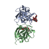
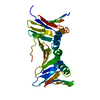
| Title | Structure of M. capricolum TrmK in complex with the natural cofactor product S-adenosyl-homocysteine (SAH) | ||||||
 Components Components | tRNA (Adenine(22)-N(1))-methyltransferase | ||||||
 Keywords Keywords | TRANSFERASE / RNA MTases / methyltransferase / m6A / transition state analogue / inhibitor / RNA binding / TrmK / RlmJ / m1A / structure. | ||||||
| Function / homology | tRNA (adenine22-N1)-methyltransferase / tRNA (adenine(22)-N1)-methyltransferase activity / tRNA methyltransferase TrmK / tRNA (adenine(22)-N(1))-methyltransferase / methylation / S-adenosyl-L-methionine-dependent methyltransferase superfamily / S-ADENOSYL-L-HOMOCYSTEINE / tRNA (Adenine(22)-N(1))-methyltransferase Function and homology information Function and homology information | ||||||
| Biological species |  Mycoplasma capricolum subsp. capricolum (bacteria) Mycoplasma capricolum subsp. capricolum (bacteria) | ||||||
| Method |  X-RAY DIFFRACTION / SYNCHROTRON / MOLECULAR REPLACEMENT / Resolution: 2.36 Å X-RAY DIFFRACTION / SYNCHROTRON / MOLECULAR REPLACEMENT / Resolution: 2.36 Å | ||||||
 Authors Authors | Oerum, S. / Catala, M. / Atdjian, C. / Brachet, F. / Ponchon, L. / Barraud, P. / Iannazzo, L. / Droogmans, L. / Braud, E. / Etheve-Quelquejeu, M. / Tisne, C. | ||||||
 Citation Citation | Journal: Rna Biol. / Year: 2019 Title: Bisubstrate analogues as structural tools to investigate m6A methyltransferase active sites. Authors: Oerum, S. / Catala, M. / Atdjian, C. / Brachet, F. / Ponchon, L. / Barraud, P. / Iannazzo, L. / Droogmans, L. / Braud, E. / Etheve-Quelquejeu, M. / Tisne, C. | ||||||
| History |
|
- Structure visualization
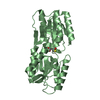
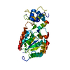
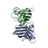
| Structure viewer | Molecule: MolmilJmol/JSmol |
|---|
- Downloads & links
-Download
| PDBx/mmCIF format | 6qe6.cif.gz | 110.7 KB | Display | PDBx/mmCIF format |
|---|---|---|---|---|
| PDB format | pdb6qe6.ent.gz | 83.6 KB | Display | PDB format |
| PDBx/mmJSON format | 6qe6.json.gz | Tree view | PDBx/mmJSON format | |
| Others |  Other downloads Other downloads |
-Validation report
| Arichive directory | https://data.pdbj.org/pub/pdb/validation_reports/qe/6qe6ftp://data.pdbj.org/pub/pdb/validation_reports/qe/6qe6 | HTTPS FTP |
|---|
-Related structure data
| Related structure data | 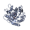 6qdxC  6qe0C 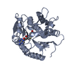 6qe5C 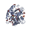 4blvS S: Starting model for refinement C: citing same article ( |
|---|---|
| Similar structure data |
-Links
 PDBj
PDBj- Assembly
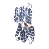
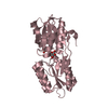
| Deposited unit | 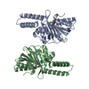
| ||||||||
|---|---|---|---|---|---|---|---|---|---|
| 1 | 
| ||||||||
| 2 | 
| ||||||||
| Unit cell |
|
-Components
| #1: Protein | Mass: 28510.572 Da / Num. of mol.: 2 Source method: isolated from a genetically manipulated source Source: (gene. exp.) Mycoplasma capricolum subsp. capricolum (bacteria)Gene: trmK, MCGM508_03695 / Production host: References: UniProt: A0A0C3A3T0, tRNA (adenine22-N1)-methyltransferase #2: Chemical |   Mass: 384.411 Da / Num. of mol.: 2 / Source method: obtained synthetically / Formula: C14H20N6O5S / Feature type: SUBJECT OF INVESTIGATION Mass: 384.411 Da / Num. of mol.: 2 / Source method: obtained synthetically / Formula: C14H20N6O5S / Feature type: SUBJECT OF INVESTIGATION#3: Water | ChemComp-HOH / |  Mass: 18.015 Da / Num. of mol.: 187 / Source method: isolated from a natural source / Formula: H2O Mass: 18.015 Da / Num. of mol.: 187 / Source method: isolated from a natural source / Formula: H2OHas protein modification | N | |
|---|
-Experimental details
-Experiment
| Experiment | Method: X-RAY DIFFRACTION / Number of used crystals: 1 |
|---|
- Sample preparation
| Crystal | Density Matthews: 2.66 Å3/Da / Density % sol: 53.75 % |
|---|---|
| Crystal grow | Temperature: 277 K / Method: vapor diffusion, sitting drop Details: 0.2 M Li2SO4, 0.1 M HEPES pH 7.5, 25% (w/v) PEG3350 |
-Data collection
| Diffraction | Mean temperature: 100 K / Serial crystal experiment: N |
|---|---|
| Diffraction source | Source: SYNCHROTRON / Site: ESRF  / Beamline: ID23-2 / Wavelength: 0.87313 Å / Beamline: ID23-2 / Wavelength: 0.87313 Å |
| Detector | Type: DECTRIS PILATUS3 X 2M / Detector: PIXEL / Date: Jun 13, 2018 |
| Radiation | Protocol: SINGLE WAVELENGTH / Monochromatic (M) / Laue (L): M / Scattering type: x-ray |
| Radiation wavelength | Wavelength: 0.87313 Å / Relative weight: 1 |
| Reflection | Resolution: 2.36→42.91 Å / Num. obs: 24468 / % possible obs: 99.75 % / Redundancy: 6.9 % / Rmerge(I) obs: 0.2719 / Net I/σ(I): 5.47 |
| Reflection shell | Resolution: 2.36→2.44 Å |
- Processing
| Software |
| ||||||||||||||||||||||||||||||||||||||||||||||||||||||||||||||||||||||
|---|---|---|---|---|---|---|---|---|---|---|---|---|---|---|---|---|---|---|---|---|---|---|---|---|---|---|---|---|---|---|---|---|---|---|---|---|---|---|---|---|---|---|---|---|---|---|---|---|---|---|---|---|---|---|---|---|---|---|---|---|---|---|---|---|---|---|---|---|---|---|---|
| Refinement | Method to determine structure: MOLECULAR REPLACEMENT Starting model: 4BLV Resolution: 2.36→42.909 Å / SU ML: 0.35 / Cross valid method: FREE R-VALUE / σ(F): 1.34 / Phase error: 27.71 / Stereochemistry target values: ML
| ||||||||||||||||||||||||||||||||||||||||||||||||||||||||||||||||||||||
| Solvent computation | Shrinkage radii: 0.9 Å / VDW probe radii: 1.11 Å / Solvent model: FLAT BULK SOLVENT MODEL | ||||||||||||||||||||||||||||||||||||||||||||||||||||||||||||||||||||||
| Refinement step | Cycle: LAST / Resolution: 2.36→42.909 Å
| ||||||||||||||||||||||||||||||||||||||||||||||||||||||||||||||||||||||
| Refine LS restraints |
| ||||||||||||||||||||||||||||||||||||||||||||||||||||||||||||||||||||||
| LS refinement shell |
|
