 Movie
Movie Controller

[English] 日本語
 Yorodumi
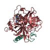
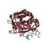
- PDB-4blv: Crystal structure of Escherichia coli 23S rRNA (A2030-N6)- methyl... -
+ Open data

- Basic information
| Entry | Database: PDB / ID: 4blv | ||||||
|---|---|---|---|---|---|---|---|
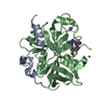
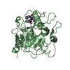
| Title | Crystal structure of Escherichia coli 23S rRNA (A2030-N6)- methyltransferase RlmJ in complex with S-adenosylmethionine (AdoMet) | ||||||
 Components Components | RIBOSOMAL RNA LARGE SUBUNIT METHYLTRANSFERASE J | ||||||
 Keywords Keywords | TRANSFERASE / N6-METHYLADENINE / ROSSMANN-LIKE FOLD / SUBDOMAIN INSERTION | ||||||
| Function / homology |  Function and homology information Function and homology information23S rRNA (adenine2030-N6)-methyltransferase / 23S rRNA (adenine(2030)-N(6))-methyltransferase activity / rRNA (adenine-N6-)-methyltransferase activity / carbon utilization / rRNA base methylation / RNA binding / cytosol Similarity search - Function | ||||||
| Biological species |  | ||||||
| Method |  X-RAY DIFFRACTION / SYNCHROTRON / MOLECULAR REPLACEMENT / Resolution: 2 Å X-RAY DIFFRACTION / SYNCHROTRON / MOLECULAR REPLACEMENT / Resolution: 2 Å | ||||||
 Authors Authors | Punekar, A.S. / Liljeruhm, J. / Shepherd, T.R. / Forster, A.C. / Selmer, M. | ||||||
 Citation Citation | Journal: Nucleic Acids Res. / Year: 2013 Title: Structural and Functional Insights Into the Molecular Mechanism of Rrna M6A Methyltransferase Rlmj. Authors: Punekar, A.S. / Liljeruhm, J. / Shepherd, T.R. / Forster, A.C. / Selmer, M. | ||||||
| History |
|
- Structure visualization
| Structure viewer | Molecule: MolmilJmol/JSmol |
|---|
- Downloads & links
-Download
| PDBx/mmCIF format | 4blv.cif.gz | 142.9 KB | Display | PDBx/mmCIF format |
|---|---|---|---|---|
| PDB format | pdb4blv.ent.gz | 111.3 KB | Display | PDB format |
| PDBx/mmJSON format | 4blv.json.gz | Tree view | PDBx/mmJSON format | |
| Others |  Other downloads Other downloads |
-Validation report
| Arichive directory | https://data.pdbj.org/pub/pdb/validation_reports/bl/4blvftp://data.pdbj.org/pub/pdb/validation_reports/bl/4blv | HTTPS FTP |
|---|
-Related structure data
| Related structure data | 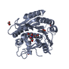 4bluSC 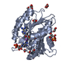 4blwC S: Starting model for refinement C: citing same article ( |
|---|---|
| Similar structure data |


-Links
 PDBj
PDBj
- Assembly
| Deposited unit | 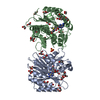
| ||||||||||||||||||||||||||||||||||||||
|---|---|---|---|---|---|---|---|---|---|---|---|---|---|---|---|---|---|---|---|---|---|---|---|---|---|---|---|---|---|---|---|---|---|---|---|---|---|---|---|
| 1 | 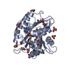
| ||||||||||||||||||||||||||||||||||||||
| 2 | 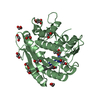
| ||||||||||||||||||||||||||||||||||||||
| Unit cell |
| ||||||||||||||||||||||||||||||||||||||
| Noncrystallographic symmetry (NCS) | NCS domain:
NCS domain segments:
NCS oper:
|
-Components
-Protein , 1 types, 2 molecules AB
| #1: Protein | Mass: 33088.871 Da / Num. of mol.: 2 Source method: isolated from a genetically manipulated source Details: SPECIFICALLY MONOMETHYLATES THE ADENINE IN POSITION 2030 OF 23S RRNA Source: (gene. exp.) References: UniProt: P37634, 23S rRNA (adenine2030-N6)-methyltransferase |
|---|
-Non-polymers , 6 types, 473 molecules 


| #2: Chemical |  Mass: 398.437 Da / Num. of mol.: 2 / Source method: obtained synthetically / Formula: C15H22N6O5S Mass: 398.437 Da / Num. of mol.: 2 / Source method: obtained synthetically / Formula: C15H22N6O5S#3: Chemical | ChemComp-GOL / |  Mass: 92.094 Da / Num. of mol.: 1 / Source method: obtained synthetically / Formula: C3H8O3 Mass: 92.094 Da / Num. of mol.: 1 / Source method: obtained synthetically / Formula: C3H8O3#4: Chemical |  Mass: 106.120 Da / Num. of mol.: 2 / Source method: obtained synthetically / Formula: C4H10O3 Mass: 106.120 Da / Num. of mol.: 2 / Source method: obtained synthetically / Formula: C4H10O3#5: Chemical | ChemComp-EDO /  Mass: 62.068 Da / Num. of mol.: 27 / Source method: obtained synthetically / Formula: C2H6O2 Mass: 62.068 Da / Num. of mol.: 27 / Source method: obtained synthetically / Formula: C2H6O2#6: Chemical |  Mass: 96.063 Da / Num. of mol.: 2 / Source method: obtained synthetically / Formula: SO4 Mass: 96.063 Da / Num. of mol.: 2 / Source method: obtained synthetically / Formula: SO4#7: Water | ChemComp-HOH / | Mass: 18.015 Da / Num. of mol.: 439 / Source method: isolated from a natural source / Formula: H2O |
|---|
-Experimental details
-Experiment
| Experiment | Method: X-RAY DIFFRACTION / Number of used crystals: 1 |
|---|
- Sample preparation
| Crystal | Density Matthews: 2.2 Å3/Da / Density % sol: 44 % / Description: NONE |
|---|---|
| Crystal grow | pH: 8.5 Details: 0.2 M SODIUM SULFATE DECAHYDRATE, 0.1 M TRIS-HCL PH 8.5 AND 30% W/V PEG 4000 |
-Data collection
| Diffraction | Mean temperature: 100 K |
|---|---|
| Diffraction source | Source: SYNCHROTRON / Site: ESRF  / Beamline: ID23-2 / Wavelength: 0.8726 / Beamline: ID23-2 / Wavelength: 0.8726 |
| Detector | Type: MARRESEARCH / Detector: CCD / Date: Oct 6, 2012 / Details: PT COATED MIRRORS |
| Radiation | Monochromator: SI (111) MONOCHROMATOR / Protocol: SINGLE WAVELENGTH / Monochromatic (M) / Laue (L): M / Scattering type: x-ray |
| Radiation wavelength | Wavelength: 0.8726 Å / Relative weight: 1 |
| Reflection | Resolution: 2→45.4 Å / Num. obs: 38776 / % possible obs: 99.8 % / Observed criterion σ(I): 2 / Redundancy: 3.8 % / Biso Wilson estimate: 18.54 Å2 / Rmerge(I) obs: 0.19 / Net I/σ(I): 8.3 |
| Reflection shell | Resolution: 2→2.1 Å / Redundancy: 3.6 % / Rmerge(I) obs: 0.75 / Mean I/σ(I) obs: 2.5 / % possible all: 99.6 |
- Processing
| Software |
| |||||||||||||||||||||||||||||||||||||||||||||||||||||||||||||||||||||||||||||||||||||||||||||||||||||||||
|---|---|---|---|---|---|---|---|---|---|---|---|---|---|---|---|---|---|---|---|---|---|---|---|---|---|---|---|---|---|---|---|---|---|---|---|---|---|---|---|---|---|---|---|---|---|---|---|---|---|---|---|---|---|---|---|---|---|---|---|---|---|---|---|---|---|---|---|---|---|---|---|---|---|---|---|---|---|---|---|---|---|---|---|---|---|---|---|---|---|---|---|---|---|---|---|---|---|---|---|---|---|---|---|---|---|---|
| Refinement | Method to determine structure: MOLECULAR REPLACEMENT Starting model: PDB ENTRY 4BLU Resolution: 2→45.39 Å / SU ML: 0.25 / σ(F): 2.01 / Phase error: 20.61 / Stereochemistry target values: ML Details: RESIDUES 53-55 IN CHAIN B ARE DISORDERED AND WERE NOT MODELED.
| |||||||||||||||||||||||||||||||||||||||||||||||||||||||||||||||||||||||||||||||||||||||||||||||||||||||||
| Solvent computation | Shrinkage radii: 0.9 Å / VDW probe radii: 1.11 Å / Solvent model: FLAT BULK SOLVENT MODEL | |||||||||||||||||||||||||||||||||||||||||||||||||||||||||||||||||||||||||||||||||||||||||||||||||||||||||
| Displacement parameters | Biso mean: 15.62 Å2 | |||||||||||||||||||||||||||||||||||||||||||||||||||||||||||||||||||||||||||||||||||||||||||||||||||||||||
| Refinement step | Cycle: LAST / Resolution: 2→45.39 Å
| |||||||||||||||||||||||||||||||||||||||||||||||||||||||||||||||||||||||||||||||||||||||||||||||||||||||||
| Refine LS restraints |
| |||||||||||||||||||||||||||||||||||||||||||||||||||||||||||||||||||||||||||||||||||||||||||||||||||||||||
| Refine LS restraints NCS |
| |||||||||||||||||||||||||||||||||||||||||||||||||||||||||||||||||||||||||||||||||||||||||||||||||||||||||
| LS refinement shell |
|
