 Movie
Movie Controller

[English] 日本語
 Yorodumi
- PDB-4io0: Crystal structure of F128A mutant of an epoxide hydrolase from Ba... -
+ Open data

- Basic information
| Entry | Database: PDB / ID: 4io0 | ||||||
|---|---|---|---|---|---|---|---|
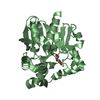
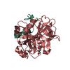
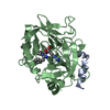
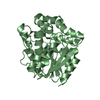
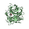
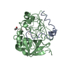
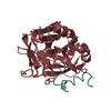
| Title | Crystal structure of F128A mutant of an epoxide hydrolase from Bacillus megaterium complexed with its product (R)-3-[1]naphthyloxy-propane-1,2-diol | ||||||
 Components Components | Soluble epoxide hydrolase | ||||||
 Keywords Keywords | HYDROLASE / a/b hydrolase fold / epoxide hydrolase | ||||||
| Function / homology |  Function and homology information Function and homology informationpyrimidine-5'-nucleotide nucleosidase / pyrimidine-5'-nucleotide nucleosidase activity Similarity search - Function | ||||||
| Biological species |  Bacillus megaterium (bacteria) Bacillus megaterium (bacteria) | ||||||
| Method |  X-RAY DIFFRACTION / SAD / Resolution: 2.9 Å X-RAY DIFFRACTION / SAD / Resolution: 2.9 Å | ||||||
 Authors Authors | Kong, X.D. / Zhou, J.H. / Xu, J.H. | ||||||
 Citation Citation | Journal: Proc.Natl.Acad.Sci.USA / Year: 2014 Title: Engineering of an epoxide hydrolase for efficient bioresolution of bulky pharmaco substrates. Authors: Kong, X.D. / Yuan, S. / Li, L. / Chen, S. / Xu, J.H. / Zhou, J.H. | ||||||
| History |
|
- Structure visualization
| Structure viewer | Molecule: MolmilJmol/JSmol |
|---|
- Downloads & links
-Download
| PDBx/mmCIF format | 4io0.cif.gz | 131.4 KB | Display | PDBx/mmCIF format |
|---|---|---|---|---|
| PDB format | pdb4io0.ent.gz | 102.5 KB | Display | PDB format |
| PDBx/mmJSON format | 4io0.json.gz | Tree view | PDBx/mmJSON format | |
| Others |  Other downloads Other downloads |
-Validation report
| Arichive directory | https://data.pdbj.org/pub/pdb/validation_reports/io/4io0ftp://data.pdbj.org/pub/pdb/validation_reports/io/4io0 | HTTPS FTP |
|---|
-Related structure data
| Related structure data | 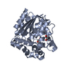 4inzC 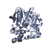 4nzzC 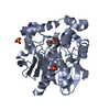 4o08C  4g00 4g02 S: Starting model for refinement C: citing same article ( |
|---|---|
| Similar structure data |


-Links
 PDBj
PDBj


- Assembly
| Deposited unit | 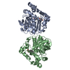
| ||||||||
|---|---|---|---|---|---|---|---|---|---|
| 1 | 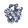
| ||||||||
| 2 | 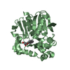
| ||||||||
| Unit cell |
|
-Components
| #1: Protein | Mass: 35210.574 Da / Num. of mol.: 2 / Mutation: F128A Source method: isolated from a genetically manipulated source Source: (gene. exp.) Bacillus megaterium (bacteria) / Strain: ECU1001 / Plasmid: pET28a / Production host: References: UniProt: G9BEX6, pyrimidine-5'-nucleotide nucleosidase #2: Chemical |   Mass: 96.063 Da / Num. of mol.: 2 / Source method: obtained synthetically / Formula: SO4 Mass: 96.063 Da / Num. of mol.: 2 / Source method: obtained synthetically / Formula: SO4#3: Chemical |   Mass: 218.248 Da / Num. of mol.: 2 / Source method: obtained synthetically / Formula: C13H14O3 Mass: 218.248 Da / Num. of mol.: 2 / Source method: obtained synthetically / Formula: C13H14O3#4: Water | ChemComp-HOH / |  Mass: 18.015 Da / Num. of mol.: 100 / Source method: isolated from a natural source / Formula: H2O Mass: 18.015 Da / Num. of mol.: 100 / Source method: isolated from a natural source / Formula: H2O |
|---|
-Experimental details
-Experiment
| Experiment | Method: X-RAY DIFFRACTION / Number of used crystals: 1 |
|---|
- Sample preparation
| Crystal | Density Matthews: 2.51 Å3/Da / Density % sol: 50.94 % |
|---|---|
| Crystal grow | Temperature: 291 K / Method: vapor diffusion, sitting drop / pH: 7.5 Details: 0.1 M Tris, 2.0 M (NH4)2SO4, 0.2 M Li2SO4, pH 7.5, VAPOR DIFFUSION, SITTING DROP, temperature 291K |
-Data collection
| Diffraction | Mean temperature: 100 K | |||||||||||||||||||||||||||||||||||||||||||||||||||||||
|---|---|---|---|---|---|---|---|---|---|---|---|---|---|---|---|---|---|---|---|---|---|---|---|---|---|---|---|---|---|---|---|---|---|---|---|---|---|---|---|---|---|---|---|---|---|---|---|---|---|---|---|---|---|---|---|---|
| Diffraction source | Source: ROTATING ANODE / Type: RIGAKU MICROMAX-007 HF / Wavelength: 1.54178 Å | |||||||||||||||||||||||||||||||||||||||||||||||||||||||
| Detector | Type: RIGAKU RAXIS IV++ / Detector: IMAGE PLATE / Date: Dec 16, 2012 / Details: mirrors | |||||||||||||||||||||||||||||||||||||||||||||||||||||||
| Radiation | Monochromator: GRAPHITE / Protocol: SINGLE WAVELENGTH / Monochromatic (M) / Laue (L): M / Scattering type: x-ray | |||||||||||||||||||||||||||||||||||||||||||||||||||||||
| Radiation wavelength | Wavelength: 1.54178 Å / Relative weight: 1 | |||||||||||||||||||||||||||||||||||||||||||||||||||||||
| Reflection | Resolution: 2.9→50 Å / Num. obs: 121961 / % possible obs: 100 % / Observed criterion σ(F): 0 / Observed criterion σ(I): -3 / Redundancy: 7.4 % / Rmerge(I) obs: 0.221 / Rsym value: 0.221 / Net I/σ(I): 8.337 | |||||||||||||||||||||||||||||||||||||||||||||||||||||||
| Reflection shell |
|
- Processing
| Software |
| ||||||||||||||||||||||||||||||||||||||||||
|---|---|---|---|---|---|---|---|---|---|---|---|---|---|---|---|---|---|---|---|---|---|---|---|---|---|---|---|---|---|---|---|---|---|---|---|---|---|---|---|---|---|---|---|
| Refinement | Method to determine structure: SAD Starting model: 4G00 4g00 Resolution: 2.9→45.25 Å / SU ML: 0.32 / σ(F): 0 / Phase error: 21.52 / Stereochemistry target values: ML
| ||||||||||||||||||||||||||||||||||||||||||
| Solvent computation | Shrinkage radii: 1.06 Å / VDW probe radii: 1.3 Å / Solvent model: FLAT BULK SOLVENT MODEL / Bsol: 22.973 Å2 / ksol: 0.342 e/Å3 | ||||||||||||||||||||||||||||||||||||||||||
| Displacement parameters |
| ||||||||||||||||||||||||||||||||||||||||||
| Refinement step | Cycle: LAST / Resolution: 2.9→45.25 Å
| ||||||||||||||||||||||||||||||||||||||||||
| Refine LS restraints |
| ||||||||||||||||||||||||||||||||||||||||||
| LS refinement shell | Refine-ID: X-RAY DIFFRACTION / Total num. of bins used: 6
|
