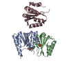
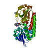
| Deposited unit | A: CMP N-GLYCOSIDASE
B: CMP N-GLYCOSIDASE
C: CMP N-GLYCOSIDASE
hetero molecules
| Theoretical mass | Number of molelcules |
|---|
| Total (without water) | 61,540 | 6 |
|---|
| Polymers | 61,255 | 3 |
|---|
| Non-polymers | 285 | 3 |
|---|
| Water | 180 | 10 |
|---|
|
|---|
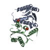
| 1 | A: CMP N-GLYCOSIDASE
B: CMP N-GLYCOSIDASE
hetero molecules
| Theoretical mass | Number of molelcules |
|---|
| Total (without water) | 41,027 | 4 |
|---|
| Polymers | 40,837 | 2 |
|---|
| Non-polymers | 190 | 2 |
|---|
| Water | 36 | 2 |
|---|
| Type | Name | Symmetry operation | Number |
|---|
| identity operation | 1_555 | x,y,z | 1 |
| Buried area | 3090 Å2 |
|---|
| ΔGint | -43 kcal/mol |
|---|
| Surface area | 12440 Å2 |
|---|
| Method | PISA |
|---|
|
|---|
| 2 | C: CMP N-GLYCOSIDASE
hetero molecules
C: CMP N-GLYCOSIDASE
hetero molecules
| Theoretical mass | Number of molelcules |
|---|
| Total (without water) | 41,027 | 4 |
|---|
| Polymers | 40,837 | 2 |
|---|
| Non-polymers | 190 | 2 |
|---|
| Water | 36 | 2 |
|---|
| Type | Name | Symmetry operation | Number |
|---|
| identity operation | 1_555 | x,y,z | 1 | | crystal symmetry operation | 2_456 | -x-1,y,-z+1 | 1 |
| Buried area | 2890 Å2 |
|---|
| ΔGint | -42 kcal/mol |
|---|
| Surface area | 11120 Å2 |
|---|
| Method | PISA |
|---|
|
|---|
| Unit cell | | Length a, b, c (Å) | 177.948, 104.006, 40.225 |
|---|
| Angle α, β, γ (deg.) | 90.000, 97.980, 90.000 |
|---|
| Int Tables number | 5 |
|---|
| Space group name H-M | C121 |
|---|
|
|---|
| Noncrystallographic symmetry (NCS) | NCS domain: | ID | Ens-ID | Details (eV) |
|---|
| 1 | 1 | chain A and (resseq 25:45 or resseq 59:108 or resseq 114:144 )| 2 | 1 | chain B and (resseq 25:45 or resseq 59:108 or resseq 114:144 )| 3 | 1 | chain C and (resseq 25:45 or resseq 59:108 or resseq 114:144 ) | | |
NCS domain segments: Ens-ID: 1 | Dom-ID | Component-ID | Beg auth comp-ID | Beg label comp-ID | End auth comp-ID | End label comp-ID | Selection details | Auth asym-ID | Label asym-ID | Auth seq-ID | Label seq-ID |
|---|
| 1 | 1 | GLNGLNASPASPchain 'A' and (resseq 25:45 or resseq 59:108 or resseq 114:144 )AA| 25 - 45 | 48 - 68 | | 1 | 2 | SERSERALAALAchain 'A' and (resseq 25:45 or resseq 59:108 or resseq 114:144 )AA| 59 - 108 | 82 - 131 | | 1 | 3 | TYRTYRLEULEUchain 'A' and (resseq 25:45 or resseq 59:108 or resseq 114:144 )AA| 114 - 144 | 137 - 167 | | 2 | 1 | GLNGLNASPASPchain 'B' and (resseq 25:45 or resseq 59:108 or resseq 114:144 )BB| 25 - 45 | 48 - 68 | | 2 | 2 | SERSERALAALAchain 'B' and (resseq 25:45 or resseq 59:108 or resseq 114:144 )BB| 59 - 108 | 82 - 131 | | 2 | 3 | TYR| TYR | | | | | | | | | | | | | | | | | | | | | | | | | | | | | | | | | | | | |
|
|---|
 Movie
Movie Controller


 Open data
 Basic information
 Components
 Keywords
 Function and homology information
 Clostridium botulinum A (bacteria)
 X-RAY DIFFRACTION /
  Authors
 Citation
 Structure visualization
 Downloads & links
 Other downloads


 PDBj
PDBj Assembly