 Movie
Movie Controller

[English] 日本語
 Yorodumi
- PDB-1n2x: Crystal Structure Analysis of TM0872, a Putative SAM-dependent Me... -
+ Open data

- Basic information
| Entry | Database: PDB / ID: 1n2x | ||||||
|---|---|---|---|---|---|---|---|
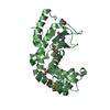
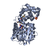
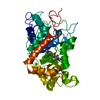
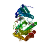
| Title | Crystal Structure Analysis of TM0872, a Putative SAM-dependent Methyltransferase, Complexed with SAM | ||||||
 Components Components | S-adenosyl-methyltransferase mraW | ||||||
 Keywords Keywords | TRANSFERASE / SAM-dependent Methyltransferase Fold / Protein-SAM Methyl Donor Complex / Structural Genomics / PSI / Protein Structure Initiative / Midwest Center for Structural Genomics / MCSG | ||||||
| Function / homology |  Function and homology information Function and homology information16S rRNA (cytosine1402-N4)-methyltransferase / rRNA (cytosine-N4-)-methyltransferase activity / rRNA base methylation / cytoplasm Similarity search - Function | ||||||
| Biological species |   Thermotoga maritima (bacteria) Thermotoga maritima (bacteria) | ||||||
| Method |  X-RAY DIFFRACTION / SYNCHROTRON / MOLECULAR REPLACEMENT / Resolution: 1.9 Å X-RAY DIFFRACTION / SYNCHROTRON / MOLECULAR REPLACEMENT / Resolution: 1.9 Å | ||||||
 Authors Authors | Miller, D.J. / Anderson, W.F. / Midwest Center for Structural Genomics (MCSG) | ||||||
 Citation Citation | Journal: Protein Sci. / Year: 2003 Title: Crystal complexes of a predicted S-adenosylmethionine-dependent methyltransferase reveal a typical AdoMet binding domain and a substrate recognition domain Authors: Miller, D.J. / Ouellette, N. / Evdokimova, E. / Savchenko, A. / Edwards, A. / Anderson, W.F. | ||||||
| History |
| ||||||
| Remark 300 | BIOMOLECULE THIS ENTRY CONTAINS THE CRYSTALLOGRAPHIC ASYMMETRIC UNIT WHICH CONSISTS OF 2 CHAINS. ...BIOMOLECULE THIS ENTRY CONTAINS THE CRYSTALLOGRAPHIC ASYMMETRIC UNIT WHICH CONSISTS OF 2 CHAINS. THE BIOLOGICAL UNIT IS A TRIMER ACCORDING TO DYNAMIC LIGHT SCATTERING DATA. |
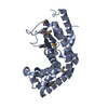
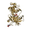
- Structure visualization
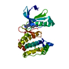
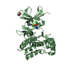
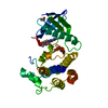
| Structure viewer | Molecule: MolmilJmol/JSmol |
|---|
- Downloads & links
-Download
| PDBx/mmCIF format | 1n2x.cif.gz | 136.3 KB | Display | PDBx/mmCIF format |
|---|---|---|---|---|
| PDB format | pdb1n2x.ent.gz | 105.3 KB | Display | PDB format |
| PDBx/mmJSON format | 1n2x.json.gz | Tree view | PDBx/mmJSON format | |
| Others |  Other downloads Other downloads |
-Validation report
| Arichive directory | https://data.pdbj.org/pub/pdb/validation_reports/n2/1n2xftp://data.pdbj.org/pub/pdb/validation_reports/n2/1n2x | HTTPS FTP |
|---|
-Related structure data
| Related structure data |  1m6ySC S: Starting model for refinement C: citing same article ( |
|---|---|
| Similar structure data | |
| Other databases |
-Links
 PDBj
PDBj
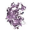
- Assembly
| Deposited unit | 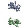
| ||||||||
|---|---|---|---|---|---|---|---|---|---|
| 1 | 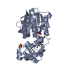
| ||||||||
| 2 | 
| ||||||||
| Unit cell |
|
-Components
| #1: Protein | Mass: 35384.977 Da / Num. of mol.: 2 Source method: isolated from a genetically manipulated source Source: (gene. exp.) Thermotoga maritima (bacteria) / Gene: TM0872 / Species (production host): Escherichia coli / Production host: References: UniProt: Q9WZX6, Transferases; Transferring one-carbon groups; Methyltransferases #2: Chemical |   Mass: 96.063 Da / Num. of mol.: 2 / Source method: obtained synthetically / Formula: SO4 Mass: 96.063 Da / Num. of mol.: 2 / Source method: obtained synthetically / Formula: SO4#3: Chemical |   Mass: 398.437 Da / Num. of mol.: 2 / Source method: obtained synthetically / Formula: C15H22N6O5S Mass: 398.437 Da / Num. of mol.: 2 / Source method: obtained synthetically / Formula: C15H22N6O5S#4: Water | ChemComp-HOH / |  Mass: 18.015 Da / Num. of mol.: 349 / Source method: isolated from a natural source / Formula: H2O Mass: 18.015 Da / Num. of mol.: 349 / Source method: isolated from a natural source / Formula: H2OHas protein modification | Y | |
|---|
-Experimental details
-Experiment
| Experiment | Method: X-RAY DIFFRACTION / Number of used crystals: 1 |
|---|
- Sample preparation
| Crystal | Density Matthews: 2.83 Å3/Da / Density % sol: 56.53 % | ||||||||||||||||||||||||||||||||||||||||||||||||||||||||||||||||||||||||||||||||||||
|---|---|---|---|---|---|---|---|---|---|---|---|---|---|---|---|---|---|---|---|---|---|---|---|---|---|---|---|---|---|---|---|---|---|---|---|---|---|---|---|---|---|---|---|---|---|---|---|---|---|---|---|---|---|---|---|---|---|---|---|---|---|---|---|---|---|---|---|---|---|---|---|---|---|---|---|---|---|---|---|---|---|---|---|---|---|
| Crystal grow | Temperature: 277 K / Method: vapor diffusion, hanging drop / pH: 6.5 Details: DROP- 50 mM NA CACODYLATE pH 6.5, 5 mM HEPES pH 7.5, 0.1 M AMMONIUM SULFATE, 9 % PEG 8000, 0.25 M NACL, 1.0 MM BME, 10 MG/ML PROTEIN. WELL- 0.1M NA CACODYLATE pH 6.5, 0.1 M AMMONIUM SULFATE, ...Details: DROP- 50 mM NA CACODYLATE pH 6.5, 5 mM HEPES pH 7.5, 0.1 M AMMONIUM SULFATE, 9 % PEG 8000, 0.25 M NACL, 1.0 MM BME, 10 MG/ML PROTEIN. WELL- 0.1M NA CACODYLATE pH 6.5, 0.1 M AMMONIUM SULFATE, 18 % PEG 8000, VAPOR DIFFUSION, HANGING DROP, temperature 277K | ||||||||||||||||||||||||||||||||||||||||||||||||||||||||||||||||||||||||||||||||||||
| Crystal grow | *PLUS Temperature: 4 ℃ | ||||||||||||||||||||||||||||||||||||||||||||||||||||||||||||||||||||||||||||||||||||
| Components of the solutions | *PLUS
|
-Data collection
| Diffraction | Mean temperature: 170 K |
|---|---|
| Diffraction source | Source: SYNCHROTRON / Site: APS  / Beamline: 5ID-B / Wavelength: 0.9785 Å / Beamline: 5ID-B / Wavelength: 0.9785 Å |
| Detector | Type: MARRESEARCH / Detector: CCD / Date: Apr 3, 2002 |
| Radiation | Monochromator: Null / Protocol: SINGLE WAVELENGTH / Monochromatic (M) / Laue (L): M / Scattering type: x-ray |
| Radiation wavelength | Wavelength: 0.9785 Å / Relative weight: 1 |
| Reflection | Resolution: 1.9→20 Å / Num. all: 63197 / Num. obs: 63100 / % possible obs: 99.8 % / Observed criterion σ(F): 0 / Observed criterion σ(I): -3 / Redundancy: 25.7 % / Biso Wilson estimate: 20.8 Å2 / Rmerge(I) obs: 0.079 / Net I/σ(I): 29 |
| Reflection shell | Resolution: 1.9→1.97 Å / Redundancy: 7 % / Rmerge(I) obs: 0.386 / Mean I/σ(I) obs: 4.65 / Num. unique all: 6264 / % possible all: 99.9 |
| Reflection | *PLUS Num. obs: 63197 / Num. measured all: 1623397 |
| Reflection shell | *PLUS % possible obs: 99.9 % / Mean I/σ(I) obs: 4.7 |
- Processing
| Software |
| ||||||||||||||||||||||||||||||||||||
|---|---|---|---|---|---|---|---|---|---|---|---|---|---|---|---|---|---|---|---|---|---|---|---|---|---|---|---|---|---|---|---|---|---|---|---|---|---|
| Refinement | Method to determine structure: MOLECULAR REPLACEMENT Starting model: PDB ENTRY 1M6Y Resolution: 1.9→19.97 Å / Isotropic thermal model: RESTRAINED / Cross valid method: THROUGHOUT / σ(F): 0 / Stereochemistry target values: Engh & Huber / Details: BULK SOLVENT MODEL USED
| ||||||||||||||||||||||||||||||||||||
| Displacement parameters | Biso mean: 40.5 Å2 | ||||||||||||||||||||||||||||||||||||
| Refine analyze |
| ||||||||||||||||||||||||||||||||||||
| Refinement step | Cycle: LAST / Resolution: 1.9→19.97 Å
| ||||||||||||||||||||||||||||||||||||
| Refine LS restraints |
| ||||||||||||||||||||||||||||||||||||
| LS refinement shell | Resolution: 1.9→1.97 Å / Rfactor Rfree error: 0.017
| ||||||||||||||||||||||||||||||||||||
| Xplor file |
| ||||||||||||||||||||||||||||||||||||
| Refinement | *PLUS Highest resolution: 1.9 Å / Lowest resolution: 20 Å | ||||||||||||||||||||||||||||||||||||
| Solvent computation | *PLUS | ||||||||||||||||||||||||||||||||||||
| Displacement parameters | *PLUS | ||||||||||||||||||||||||||||||||||||
| Refine LS restraints | *PLUS
|
