 Movie
Movie Controller

[English] 日本語
 Yorodumi
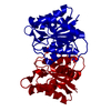
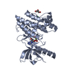
- PDB-4jem: Crystal structure of MilB complexed with cytidine 5'-monophosphate -
+ Open data

- Basic information
| Entry | Database: PDB / ID: 4jem | ||||||
|---|---|---|---|---|---|---|---|
| Title | Crystal structure of MilB complexed with cytidine 5'-monophosphate | ||||||
 Components Components | CMP/hydroxymethyl CMP hydrolase | ||||||
 Keywords Keywords | HYDROLASE / CMP N-glycosidase | ||||||
| Function / homology | Nucleoside 2-deoxyribosyltransferase / Nucleoside 2-deoxyribosyltransferase / Rossmann fold - #450 / hydrolase activity / Rossmann fold / 3-Layer(aba) Sandwich / Alpha Beta / CYTIDINE-5'-MONOPHOSPHATE / CMP/hydroxymethyl CMP hydrolase Function and homology information Function and homology information | ||||||
| Biological species |  Streptomyces rimofaciens (bacteria) Streptomyces rimofaciens (bacteria) | ||||||
| Method |  X-RAY DIFFRACTION / SYNCHROTRON / FOURIER SYNTHESIS / Resolution: 1.553 Å X-RAY DIFFRACTION / SYNCHROTRON / FOURIER SYNTHESIS / Resolution: 1.553 Å | ||||||
 Authors Authors | Sikowitz, M.D. / Cooper, L.E. / Begley, T.P. / Kaminski, P.A. / Ealick, S.E. | ||||||
 Citation Citation | Journal: Biochemistry / Year: 2013 Title: Reversal of the substrate specificity of CMP N-glycosidase to dCMP. Authors: Sikowitz, M.D. / Cooper, L.E. / Begley, T.P. / Kaminski, P.A. / Ealick, S.E. | ||||||
| History |
|
- Structure visualization
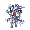
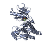
| Structure viewer | Molecule: MolmilJmol/JSmol |
|---|
- Downloads & links
-Download
| PDBx/mmCIF format | 4jem.cif.gz | 77 KB | Display | PDBx/mmCIF format |
|---|---|---|---|---|
| PDB format | pdb4jem.ent.gz | 56.2 KB | Display | PDB format |
| PDBx/mmJSON format | 4jem.json.gz | Tree view | PDBx/mmJSON format | |
| Others |  Other downloads Other downloads |
-Validation report
| Arichive directory | https://data.pdbj.org/pub/pdb/validation_reports/je/4jemftp://data.pdbj.org/pub/pdb/validation_reports/je/4jem | HTTPS FTP |
|---|
-Related structure data
| Related structure data |  4jelSC  4jenC S: Starting model for refinement C: citing same article ( |
|---|---|
| Similar structure data |


-Links
 PDBj
PDBj- Assembly
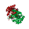
| Deposited unit | 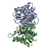
| ||||||||
|---|---|---|---|---|---|---|---|---|---|
| 1 |
| ||||||||
| Unit cell |
|
-Components
| #1: Protein | Mass: 18381.641 Da / Num. of mol.: 2 Source method: isolated from a genetically manipulated source Source: (gene. exp.) Streptomyces rimofaciens (bacteria) / Gene: MilB / Plasmid: pET28a / Production host: #2: Chemical |   Mass: 323.197 Da / Num. of mol.: 2 / Source method: obtained synthetically / Formula: C9H14N3O8P Mass: 323.197 Da / Num. of mol.: 2 / Source method: obtained synthetically / Formula: C9H14N3O8P#3: Water | ChemComp-HOH / |  Mass: 18.015 Da / Num. of mol.: 194 / Source method: isolated from a natural source / Formula: H2O Mass: 18.015 Da / Num. of mol.: 194 / Source method: isolated from a natural source / Formula: H2O |
|---|
-Experimental details
-Experiment
| Experiment | Method: X-RAY DIFFRACTION / Number of used crystals: 1 |
|---|
- Sample preparation
| Crystal | Density Matthews: 2.17 Å3/Da / Density % sol: 43.28 % |
|---|---|
| Crystal grow | Temperature: 291 K / Method: vapor diffusion, hanging drop / pH: 7.5 Details: 0.2 M magnesium chloride, 18% PEG 8000, 0.1 M Tris, pH 7.5, VAPOR DIFFUSION, HANGING DROP, temperature 291K |
-Data collection
| Diffraction | Mean temperature: 100 K | ||||||||||||||||||||||||||||
|---|---|---|---|---|---|---|---|---|---|---|---|---|---|---|---|---|---|---|---|---|---|---|---|---|---|---|---|---|---|
| Diffraction source | Source: SYNCHROTRON / Site: APS  / Beamline: 24-ID-C / Wavelength: 0.97918 Å / Beamline: 24-ID-C / Wavelength: 0.97918 Å | ||||||||||||||||||||||||||||
| Detector | Type: ADSC QUANTUM 315 / Detector: CCD / Date: Apr 8, 2011 | ||||||||||||||||||||||||||||
| Radiation | Monochromator: cryocooled Si(111) double crystal / Protocol: SINGLE WAVELENGTH / Monochromatic (M) / Laue (L): M / Scattering type: x-ray | ||||||||||||||||||||||||||||
| Radiation wavelength | Wavelength: 0.97918 Å / Relative weight: 1 | ||||||||||||||||||||||||||||
| Reflection | Resolution: 1.55→50 Å / Num. all: 44996 / Num. obs: 44996 / % possible obs: 99.8 % / Observed criterion σ(I): -3 / Biso Wilson estimate: 15.64 Å2 / Rsym value: 0.057 / Net I/σ(I): 25.1 | ||||||||||||||||||||||||||||
| Reflection shell |
|
- Processing
| Software |
| |||||||||||||||||||||||||||||||||||||||||||||||||||||||||||||||||||||||||||||||||||||||||||||||||||||||||||||||||||||||
|---|---|---|---|---|---|---|---|---|---|---|---|---|---|---|---|---|---|---|---|---|---|---|---|---|---|---|---|---|---|---|---|---|---|---|---|---|---|---|---|---|---|---|---|---|---|---|---|---|---|---|---|---|---|---|---|---|---|---|---|---|---|---|---|---|---|---|---|---|---|---|---|---|---|---|---|---|---|---|---|---|---|---|---|---|---|---|---|---|---|---|---|---|---|---|---|---|---|---|---|---|---|---|---|---|---|---|---|---|---|---|---|---|---|---|---|---|---|---|---|---|
| Refinement | Method to determine structure: FOURIER SYNTHESIS Starting model: 4JEL Resolution: 1.553→40.858 Å / Occupancy max: 1 / Occupancy min: 1 / FOM work R set: 0.87 / SU ML: 0.15 / σ(F): 1.34 / Phase error: 20.54 / Stereochemistry target values: ML
| |||||||||||||||||||||||||||||||||||||||||||||||||||||||||||||||||||||||||||||||||||||||||||||||||||||||||||||||||||||||
| Solvent computation | Shrinkage radii: 0.9 Å / VDW probe radii: 1.11 Å / Solvent model: FLAT BULK SOLVENT MODEL | |||||||||||||||||||||||||||||||||||||||||||||||||||||||||||||||||||||||||||||||||||||||||||||||||||||||||||||||||||||||
| Displacement parameters | Biso max: 47.53 Å2 / Biso mean: 17.0707 Å2 / Biso min: 7.02 Å2 | |||||||||||||||||||||||||||||||||||||||||||||||||||||||||||||||||||||||||||||||||||||||||||||||||||||||||||||||||||||||
| Refinement step | Cycle: LAST / Resolution: 1.553→40.858 Å
| |||||||||||||||||||||||||||||||||||||||||||||||||||||||||||||||||||||||||||||||||||||||||||||||||||||||||||||||||||||||
| Refine LS restraints |
| |||||||||||||||||||||||||||||||||||||||||||||||||||||||||||||||||||||||||||||||||||||||||||||||||||||||||||||||||||||||
| LS refinement shell | Refine-ID: X-RAY DIFFRACTION / Total num. of bins used: 16
|
