[English] 日本語
Yorodumi
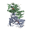
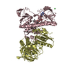
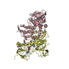
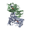
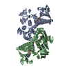
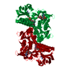
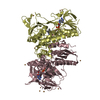
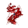
- PDB-4k7z: Crystal structure of the C136(42)A/C141(47)A double mutant of Tn5... -

+
Open data


ID or keywords:

Loading...

-
Basic information

Entry
Database: PDB / ID: 4k7z
TitleCrystal structure of the C136(42)A/C141(47)A double mutant of Tn501 MerA in complex with NADP and Hg2+
ComponentsMercuric reductase
KeywordsOXIDOREDUCTASE / mercuric ion reductase / flavoenzyme / mercuric ion complex / NADPH complex / ternary complex / reduced form
Function / homology
Function and homology information


mercury(II) reductase / mercury (II) reductase (NADP+) activity / oxidoreductase activity, acting on a sulfur group of donors, NAD(P) as acceptor / detoxification of mercury ion / mercury ion binding / NADP binding / flavin adenine dinucleotide binding
Similarity search - Function
Mercury(II) reductase / Heavy-metal-associated, conserved site / Heavy-metal-associated domain. / Heavy-metal-associated domain / Heavy metal-associated domain superfamily / Heavy-metal-associated domain profile. / Heavy metal-associated domain, HMA / Pyridine nucleotide-disulphide oxidoreductase, class I / FAD/NAD-linked reductase, C-terminal dimerisation domain / Pyridine nucleotide-disulphide oxidoreductase, class I, active site ...Mercury(II) reductase / Heavy-metal-associated, conserved site / Heavy-metal-associated domain. / Heavy-metal-associated domain / Heavy metal-associated domain superfamily / Heavy-metal-associated domain profile. / Heavy metal-associated domain, HMA / Pyridine nucleotide-disulphide oxidoreductase, class I / FAD/NAD-linked reductase, C-terminal dimerisation domain / Pyridine nucleotide-disulphide oxidoreductase, class I, active site / Pyridine nucleotide-disulphide oxidoreductases class-I active site. / Pyridine nucleotide-disulphide oxidoreductase, dimerisation domain / Pyridine nucleotide-disulphide oxidoreductase, dimerisation domain / FAD/NAD-linked reductase, dimerisation domain superfamily / FAD/NAD(P)-binding domain / Pyridine nucleotide-disulphide oxidoreductase / Enolase-like; domain 1 / FAD/NAD(P)-binding domain / FAD/NAD(P)-binding domain / 3-Layer(bba) Sandwich / FAD/NAD(P)-binding domain superfamily / 2-Layer Sandwich / Alpha Beta
Similarity search - Domain/homology
FLAVIN-ADENINE DINUCLEOTIDE / : / NADP NICOTINAMIDE-ADENINE-DINUCLEOTIDE PHOSPHATE / Mercuric reductase
Similarity search - Component
Biological speciesPseudomonas aeruginosa (bacteria)
MethodX-RAY DIFFRACTION / SYNCHROTRON / MOLECULAR REPLACEMENT / Resolution: 1.5 Å
AuthorsDong, A. / Falkowaski, M. / Malone, M. / Miller, S.M. / Pai, E.F.
CitationJournal: To be Published
Title: Crystal structure of the C136(42)A/C141(47)A double mutant of Tn501 MerA in complex with NADP and Hg2+
Authors: Dong, A. / Falkowski, M. / Malone, M. / Miller, S.M. / Pai, E.F.
History
DepositionApr 17, 2013Deposition site: RCSB / Processing site: RCSB
Revision 1.0May 15, 2013Provider: repository / Type: Initial release
Revision 1.1Sep 20, 2023Group: Data collection / Database references ...Data collection / Database references / Derived calculations / Refinement description
Category: chem_comp_atom / chem_comp_bond ...chem_comp_atom / chem_comp_bond / database_2 / pdbx_initial_refinement_model / struct_ref_seq_dif / struct_site
Item: _database_2.pdbx_DOI / _database_2.pdbx_database_accession ..._database_2.pdbx_DOI / _database_2.pdbx_database_accession / _struct_ref_seq_dif.details / _struct_site.pdbx_auth_asym_id / _struct_site.pdbx_auth_comp_id / _struct_site.pdbx_auth_seq_id

-
Structure visualization

Structure viewerMolecule:
MolmilJmol/JSmol

Downloads & links

-
Assembly

Deposited unit
A: Mercuric reductase
hetero molecules


Theoretical massNumber of molelcules
Total (without water)51,45626
Polymers49,1621
Non-polymers2,29425
Water10,124562
1
A: Mercuric reductase
hetero molecules

A: Mercuric reductase
hetero molecules


Theoretical massNumber of molelcules
Total (without water)102,91152
Polymers98,3232
Non-polymers4,58850
Water362
TypeNameSymmetry operationNumber
identity operation1_555x,y,z1
crystal symmetry operation8_555-y,-x,-z+1/21
Buried area15780 Å2
ΔGint-139 kcal/mol
Surface area34220 Å2
MethodPISA
Unit cell
Length a, b, c (Å)86.584, 86.584, 137.370
Angle α, β, γ (deg.)90.00, 90.00, 90.00
Int Tables number96
Space group name H-MP43212
Components on special symmetry positions
IDModelComponents
11A-1130-

HOH

-
Components

-
Protein , 1 types, 1 molecules A

#1: Protein Mercuric reductase / Hg(II) reductase


Mass: 49161.680 Da / Num. of mol.: 1 / Fragment: unp residues 96-561 / Mutation: C42A, C47A
Source method: isolated from a genetically manipulated source
Source: (gene. exp.) Pseudomonas aeruginosa (bacteria) / Gene: merA / Plasmid: pET3d:cmerA / Production host: Escherichia coli (E. coli) / Strain (production host): BL21-(DE3)pLysS / References: UniProt: P00392, mercury(II) reductase

-
Non-polymers , 7 types, 587 molecules

#2: Chemical ChemComp-FAD / FLAVIN-ADENINE DINUCLEOTIDE


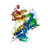
Mass: 785.550 Da / Num. of mol.: 1 / Source method: obtained synthetically / Formula: C27H33N9O15P2 / Comment: FAD*YM
#3: Chemical ChemComp-NAP / NADP NICOTINAMIDE-ADENINE-DINUCLEOTIDE PHOSPHATE / 2'-MONOPHOSPHOADENOSINE 5'-DIPHOSPHORIBOSE


Mass: 743.405 Da / Num. of mol.: 1 / Source method: obtained synthetically / Formula: C21H28N7O17P3
#4: Chemical ChemComp-HG / MERCURY (II) ION


Mass: 200.590 Da / Num. of mol.: 1 / Source method: obtained synthetically / Formula: Hg
#5: Chemical ChemComp-SO4 / SULFATE ION


Mass: 96.063 Da / Num. of mol.: 3 / Source method: obtained synthetically / Formula: SO4
#6: Chemical ChemComp-GOL / GLYCEROL / GLYCERIN / PROPANE-1,2,3-TRIOL


Mass: 92.094 Da / Num. of mol.: 3 / Source method: obtained synthetically / Formula: C3H8O3
#7: Chemical
ChemComp-UNX / UNKNOWN ATOM OR ION


Num. of mol.: 16 / Source method: obtained synthetically
#8: Water ChemComp-HOH / water


Mass: 18.015 Da / Num. of mol.: 562 / Source method: isolated from a natural source / Formula: H2O

-
Experimental details

-
Experiment

ExperimentMethod: X-RAY DIFFRACTION / Number of used crystals: 1

-
Sample preparation

CrystalDensity Matthews: 2.62 Å3/Da / Density % sol: 53.03 %
Crystal growTemperature: 295 K / Method: vapor diffusion, hanging drop / pH: 9.4
Details: 2.0M AMMONIA Sulfate, 0.1M Tris pH9.4, vapor diffusion hanging drop, temperature 295K, VAPOR DIFFUSION, HANGING DROP

-
Data collection

DiffractionMean temperature: 100 K
Diffraction sourceSource: SYNCHROTRON / Site: APS / Beamline: 14-BM-C / Wavelength: 0.9 Å
DetectorType: ADSC QUANTUM 4 / Detector: CCD / Date: Nov 2, 2002
RadiationProtocol: SINGLE WAVELENGTH / Monochromatic (M) / Laue (L): M / Scattering type: x-ray
Radiation wavelengthWavelength: 0.9 Å / Relative weight: 1
ReflectionResolution: 1.5→80 Å / Num. obs: 83679 / % possible obs: 99.3 % / Observed criterion σ(F): 0 / Observed criterion σ(I): 0 / Redundancy: 7 % / Biso Wilson estimate: 15.6 Å2 / Rmerge(I) obs: 0.05 / Rsym value: 0.05 / Net I/σ(I): 30
Reflection shellResolution: 1.5→1.53 Å / Rmerge(I) obs: 0.56 / Mean I/σ(I) obs: 1.74 / Num. unique all: 3831 / % possible all: 92.4

-
Processing

Software
NameVersionClassificationNB
REFMAC5.7.0032refinement
PDB_EXTRACT3.11data extraction
DENZOdata reduction
SCALEPACKdata scaling
CNSphasing
Coot0.6.2model building
RefinementMethod to determine structure: MOLECULAR REPLACEMENT
Starting model: 3GRS
Resolution: 1.5→26.87 Å / Cor.coef. Fo:Fc: 0.964 / Cor.coef. Fo:Fc free: 0.951 / Occupancy max: 1 / Occupancy min: 0.3 / SU B: 1.198 / SU ML: 0.045 / Cross valid method: THROUGHOUT / σ(F): 0 / ESU R: 0.07 / ESU R Free: 0.071 / Stereochemistry target values: MAXIMUM LIKELIHOOD
RfactorNum. reflection% reflectionSelection details
Rfree0.2064 1658 2 %RANDOM
Rwork0.1812 ---
obs0.1817 83623 99.3 %-
Solvent computationIon probe radii: 0.8 Å / Shrinkage radii: 0.8 Å / VDW probe radii: 1.2 Å / Solvent model: MASK
Displacement parametersBiso max: 78.11 Å2 / Biso mean: 21.3006 Å2 / Biso min: 11.6 Å2
Baniso -1Baniso -2Baniso -3
1-0.44 Å20 Å20 Å2
2--0.44 Å2-0 Å2
3----0.89 Å2
Refinement stepCycle: LAST / Resolution: 1.5→26.87 Å
ProteinNucleic acidLigandSolventTotal
Num. atoms3413 0 151 562 4126
Refine LS restraints
Refine-IDTypeDev idealDev ideal targetNumber
X-RAY DIFFRACTIONr_bond_refined_d0.0060.0193699
X-RAY DIFFRACTIONr_bond_other_d0.0010.023514
X-RAY DIFFRACTIONr_angle_refined_deg1.2992.0025080
X-RAY DIFFRACTIONr_angle_other_deg0.73738071
X-RAY DIFFRACTIONr_dihedral_angle_1_deg5.5925496
X-RAY DIFFRACTIONr_dihedral_angle_2_deg37.01824.384146
X-RAY DIFFRACTIONr_dihedral_angle_3_deg11.52115563
X-RAY DIFFRACTIONr_dihedral_angle_4_deg14.4271526
X-RAY DIFFRACTIONr_chiral_restr0.0690.2611
X-RAY DIFFRACTIONr_gen_planes_refined0.0050.0214227
X-RAY DIFFRACTIONr_gen_planes_other0.0010.02794
X-RAY DIFFRACTIONr_mcbond_it0.8671.8821900
X-RAY DIFFRACTIONr_mcbond_other0.8641.8811899
X-RAY DIFFRACTIONr_mcangle_it1.4962.8232382
LS refinement shellResolution: 1.499→1.538 Å / Total num. of bins used: 20
RfactorNum. reflection% reflection
Rfree0.292 116 -
Rwork0.288 5637 -
all-5753 -
obs--93.76 %

+
About Yorodumi

-
News

-
Feb 9, 2022. New format data for meta-information of EMDB entries

New format data for meta-information of EMDB entries

  • Version 3 of the EMDB header file is now the official format.
  • The previous official version 1.9 will be removed from the archive.

Related info.:EMDB header

External links:wwPDB to switch to version 3 of the EMDB data model

-
Aug 12, 2020. Covid-19 info

Covid-19 info

URL: https://pdbjlvh1.pdbj.org/emnavi/covid19.php

New page: Covid-19 featured information page in EM Navigator.

Related info.:Covid-19 info / Mar 5, 2020. Novel coronavirus structure data

+
Mar 5, 2020. Novel coronavirus structure data

Novel coronavirus structure data

Related info.:Yorodumi Speices / Aug 12, 2020. Covid-19 info

External links:COVID-19 featured content - PDBj / Molecule of the Month (242):Coronavirus Proteases

+
Jan 31, 2019. EMDB accession codes are about to change! (news from PDBe EMDB page)

EMDB accession codes are about to change! (news from PDBe EMDB page)

  • The allocation of 4 digits for EMDB accession codes will soon come to an end. Whilst these codes will remain in use, new EMDB accession codes will include an additional digit and will expand incrementally as the available range of codes is exhausted. The current 4-digit format prefixed with “EMD-” (i.e. EMD-XXXX) will advance to a 5-digit format (i.e. EMD-XXXXX), and so on. It is currently estimated that the 4-digit codes will be depleted around Spring 2019, at which point the 5-digit format will come into force.
  • The EM Navigator/Yorodumi systems omit the EMD- prefix.

Related info.:Q: What is EMD? / ID/Accession-code notation in Yorodumi/EM Navigator

External links:EMDB Accession Codes are Changing Soon! / Contact to PDBj

+
Jul 12, 2017. Major update of PDB

Major update of PDB

  • wwPDB released updated PDB data conforming to the new PDBx/mmCIF dictionary.
  • This is a major update changing the version number from 4 to 5, and with Remediation, in which all the entries are updated.
  • In this update, many items about electron microscopy experimental information are reorganized (e.g. em_software).
  • Now, EM Navigator and Yorodumi are based on the updated data.

External links:wwPDB Remediation / Enriched Model Files Conforming to OneDep Data Standards Now Available in the PDB FTP Archive

-
Yorodumi

Thousand views of thousand structures

  • Yorodumi is a browser for structure data from EMDB, PDB, SASBDB, etc.
  • This page is also the successor to EM Navigator detail page, and also detail information page/front-end page for Omokage search.
  • The word "yorodu" (or yorozu) is an old Japanese word meaning "ten thousand". "mi" (miru) is to see.

Related info.:EMDB / PDB / SASBDB / Comparison of 3 databanks / Yorodumi Search / Aug 31, 2016. New EM Navigator & Yorodumi / Yorodumi Papers / Jmol/JSmol / Function and homology information / Changes in new EM Navigator and Yorodumi

Read more