Component-ID: _ / Ens-ID: 1 / Beg auth comp-ID: ALA / Beg label comp-ID: ALA / End auth comp-ID: TYR / End label comp-ID: TYR / Refine code: _ / Auth seq-ID: 1 - 452 / Label seq-ID: 1 - 422
Dom-ID
Auth asym-ID
Label asym-ID
1
A
A
2
B
B
-
Components
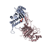
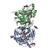
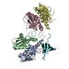
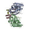
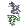
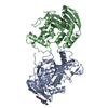
#1: Protein
Lysosomalprotectiveprotein / Carboxypeptidase C / Carboxypeptidase L / Cathepsin A / Protective protein cathepsin A / PPCA / ...Carboxypeptidase C / Carboxypeptidase L / Cathepsin A / Protective protein cathepsin A / PPCA / Protective protein for beta-galactosidase
Mass: 48655.582 Da / Num. of mol.: 2 Source method: isolated from a genetically manipulated source Details: Selected with blasticidin / Source: (gene. exp.) Homo sapiens (human) / Gene: CTSA, PPGB / Plasmid: pIB/V5-His-TOPO TA / Production host: Trichoplusia ni (cabbage looper) / Strain (production host): HI-FIVE / References: UniProt: P10619, carboxypeptidase C
Type: D-saccharide, beta linking / Mass: 221.208 Da / Num. of mol.: 3 Source method: isolated from a genetically manipulated source Formula: C8H15NO6
Identifier
Type
Program
DGlcpNAcb
CONDENSED IUPAC CARBOHYDRATE SYMBOL
GMML 1.0
N-acetyl-b-D-glucopyranosamine
COMMON NAME
GMML 1.0
b-D-GlcpNAc
IUPAC CARBOHYDRATE SYMBOL
PDB-CARE 1.0
GlcNAc
SNFG CARBOHYDRATE SYMBOL
GMML 1.0
Has protein modification
Y
Sequence details
AN UNIDENTIFIED PROTEASE CONVERTED THE ZYMOGEN INTO ACTIVE ENZYME. PUTATIVE TERMINI ASSUME ...AN UNIDENTIFIED PROTEASE CONVERTED THE ZYMOGEN INTO ACTIVE ENZYME. PUTATIVE TERMINI ASSUME PROTEOLYTIC REMOVAL OF 263-292, CONSISTENT WITH MASS SPECTROMETRY AND N-TERMINAL SEQUENCING DATA.
-
Experimental details
-
Experiment
Experiment
Method: X-RAY DIFFRACTION / Number of used crystals: 1
-
Sample preparation
Crystal
Density Matthews: 2.68 Å3/Da / Density % sol: 54.08 %
Crystal grow
Temperature: 293 K / Method: vapor diffusion, sitting drop / pH: 7 Details: 10% PEG 3350, 0.1M ammonium tartrate, pH 7.0, VAPOR DIFFUSION, SITTING DROP, temperature 293K
 Movie
Movie Controller


 Open data
 Basic information
 Components
 Keywords
 Function and homology information
 Homo sapiens (human)
 X-RAY DIFFRACTION /
  Authors
 Citation
 Structure visualization
 Downloads & links
 Other downloads


 PDBj
PDBj


 Assembly
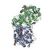
 Trichoplusia ni (cabbage looper) / Strain (production host): HI-FIVE / References: UniProt: P10619, carboxypeptidase C
 Type: D-saccharide, beta linking / Mass: 221.208 Da / Num. of mol.: 3
 Sample preparation
 / Beamline: 24-ID-C / Wavelength: 0.9792 Å
 Processing