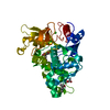
| Deposited unit | A: Lipase 46 kDa form
B: Lipase 46 kDa form
hetero molecules
| Theoretical mass | Number of molelcules |
|---|
| Total (without water) | 96,926 | 6 |
|---|
| Polymers | 96,715 | 2 |
|---|
| Non-polymers | 211 | 4 |
|---|
| Water | 468 | 26 |
|---|
|
|---|
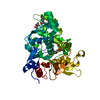
| 1 | A: Lipase 46 kDa form
hetero molecules
| Theoretical mass | Number of molelcules |
|---|
| Total (without water) | 48,463 | 3 |
|---|
| Polymers | 48,358 | 1 |
|---|
| Non-polymers | 105 | 2 |
|---|
| Water | 18 | 1 |
|---|
| Type | Name | Symmetry operation | Number |
|---|
| identity operation | 1_555 | x,y,z | 1 |
|
|---|
| 2 | B: Lipase 46 kDa form
hetero molecules
| Theoretical mass | Number of molelcules |
|---|
| Total (without water) | 48,463 | 3 |
|---|
| Polymers | 48,358 | 1 |
|---|
| Non-polymers | 105 | 2 |
|---|
| Water | 18 | 1 |
|---|
| Type | Name | Symmetry operation | Number |
|---|
| identity operation | 1_555 | x,y,z | 1 |
|
|---|
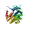
| 3 |
- Idetical with deposited unit
- defined by software
| Type | Name | Symmetry operation | Number |
|---|
| identity operation | 1_555 | x,y,z | 1 |
| Buried area | 2820 Å2 |
|---|
| ΔGint | -56 kcal/mol |
|---|
| Surface area | 32170 Å2 |
|---|
| Method | PISA, PQS |
|---|
|
|---|
| Unit cell | | Length a, b, c (Å) | 73.310, 77.960, 169.810 |
|---|
| Angle α, β, γ (deg.) | 90.00, 90.00, 90.00 |
|---|
| Int Tables number | 19 |
|---|
| Space group name H-M | P212121 |
|---|
|
|---|
| Noncrystallographic symmetry (NCS) | NCS domain: | ID | Ens-ID | Details (eV) |
|---|
| 1 | 1 | A| 2 | 1 | B| 3 | 1 | A| 4 | 1 | B| 5 | 1 | A| 6 | 1 | B| 7 | 1 | A| 8 | 1 | B| 9 | 1 | A| 10 | 1 | B| 11 | 1 | A| 12 | 1 | B| 13 | 1 | A| 14 | 1 | B| 15 | 1 | A| 16 | 1 | B| 17 | 1 | A| 18 | 1 | B| 19 | 1 | A| 20 | 1 | B| 21 | 1 | A| 22 | 1 | B| 23 | 1 | A| 24 | 1 | B | | | | | | | | | | | | | | | | | | | | | | | |
NCS domain segments: Ens-ID: 1 / Refine code: 4 | Dom-ID | Component-ID | Beg auth comp-ID | Beg label comp-ID | End auth comp-ID | End label comp-ID | Auth asym-ID | Label asym-ID | Auth seq-ID | Label seq-ID |
|---|
| 1 | 1 | PHEPHEALAALAAA| 29 - 35 | 64 - 70 | | 2 | 1 | PHEPHEALAALABB| 29 - 35 | 64 - 70 | | 3 | 2 | ALAALAPROPROAA| 7 - 11 | 42 - 46 | | 4 | 2 | ALAALAPROPROBB| 7 - 11 | 42 - 46 | | 5 | 3 | TYRTYRGLYGLYAA| 40 - 84 | 75 - 119 | | 6 | 3 | TYRTYRGLYGLYBB| 40 - 84 | 75 - 119 | | 7 | 4 | GLNGLNLYSLYSAA| 201 - 221 | 236 - 256 | | 8 | 4 | GLNGLNLYSLYSBB| 201 - 221 | 236 - 256 | | 9 | 5 | GLYGLYSERSERAA| 283 - 326 | 318 - 361 | | 10 | 5 | GLYGLYSERSERBB| 283 - 326 | 318 - 361 | | 11 | 6 | LYSLYSSERSERAA| 329 - 337 | 364 - 372 | | 12 | 6 | LYSLYSSERSERBB| 329 - 337 | 364 - 372 | | 13 | 7 | HISHISSERSERA| A | | | | | | | | | | | | | | | | | | | | | | | | | | | | | | | | | | | | | | | | | | | | | | | | | | | | | | | | | | | | | | | | | | | | | | | | | | | | | |
|
|---|
 Movie
Movie Controller


 Open data
 Basic information
 Components
 Keywords
 Function and homology information
 Staphylococcus hyicus (bacteria)
 X-RAY DIFFRACTION /
  Authors
 Citation
 Structure visualization
 Downloads & links
 Other downloads


 PDBj
PDBj Assembly

