 Movie
Movie Controller


+ Open data

- Basic information
| Entry | Database: PDB / ID: 6eyy | ||||||
|---|---|---|---|---|---|---|---|
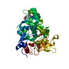
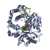
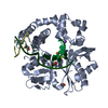
| Title | Anti-CRISPR AcrIIa6 cubic form | ||||||
 Components Components | AcrIIa6 | ||||||
 Keywords Keywords | VIRAL PROTEIN / Anti-CRISPR / HTH fold / DNA binding | ||||||
| Function / homology | : / Acr-like protein Function and homology information Function and homology information | ||||||
| Biological species |  Streptococcus phage 73 (virus) Streptococcus phage 73 (virus) | ||||||
| Method |  X-RAY DIFFRACTION / SYNCHROTRON / MOLECULAR REPLACEMENT / Resolution: 2.5 Å X-RAY DIFFRACTION / SYNCHROTRON / MOLECULAR REPLACEMENT / Resolution: 2.5 Å | ||||||
 Authors Authors | Cambillau, C. / Amigues, B. / Moineau, S. | ||||||
 Citation Citation | Journal: Nat Commun / Year: 2018 Title: Widespread anti-CRISPR proteins in virulent bacteriophages inhibit a range of Cas9 proteins. Authors: Hynes, A.P. / Rousseau, G.M. / Agudelo, D. / Goulet, A. / Amigues, B. / Loehr, J. / Romero, D.A. / Fremaux, C. / Horvath, P. / Doyon, Y. / Cambillau, C. / Moineau, S. | ||||||
| History |
|
- Structure visualization
| Structure viewer | Molecule: MolmilJmol/JSmol |
|---|
- Downloads & links
-Download
| PDBx/mmCIF format | 6eyy.cif.gz | 164.4 KB | Display | PDBx/mmCIF format |
|---|---|---|---|---|
| PDB format | pdb6eyy.ent.gz | 132.9 KB | Display | PDB format |
| PDBx/mmJSON format | 6eyy.json.gz | Tree view | PDBx/mmJSON format | |
| Others |  Other downloads Other downloads |
-Validation report
| Arichive directory | https://data.pdbj.org/pub/pdb/validation_reports/ey/6eyyftp://data.pdbj.org/pub/pdb/validation_reports/ey/6eyy | HTTPS FTP |
|---|
-Related structure data


-Links
 PDBj
PDBj- Assembly
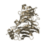
| Deposited unit | 
| ||||||||
|---|---|---|---|---|---|---|---|---|---|
| 1 |
| ||||||||
| Unit cell |
|
-Components
| #1: Protein | Mass: 21464.391 Da / Num. of mol.: 2 Source method: isolated from a genetically manipulated source Source: (gene. exp.) Streptococcus phage 73 (virus) / Variant: Steptococcus thermophilus phage D1811 / Production host:  #2: Water | ChemComp-HOH / |  Mass: 18.015 Da / Num. of mol.: 287 / Source method: isolated from a natural source / Formula: H2O Mass: 18.015 Da / Num. of mol.: 287 / Source method: isolated from a natural source / Formula: H2OSource details | Streptococcus thermophilus phage D1176 | |
|---|
-Experimental details
-Experiment
| Experiment | Method: X-RAY DIFFRACTION / Number of used crystals: 1 |
|---|
- Sample preparation
| Crystal | Density Matthews: 5.23 Å3/Da / Density % sol: 76.46 % |
|---|---|
| Crystal grow | Temperature: 293 K / Method: vapor diffusion, sitting drop Details: 0.2M Magnesium chloride, 0.1M tris pH6.5-7.5, 5-15%(w/v) PEG 8000. PH range: 6.5 - 7.5 |
-Data collection
| Diffraction | Mean temperature: 100 K |
|---|---|
| Diffraction source | Source: SYNCHROTRON / Site: SOLEIL  / Beamline: PROXIMA 1 / Wavelength: 0.9789 Å / Beamline: PROXIMA 1 / Wavelength: 0.9789 Å |
| Detector | Type: DECTRIS PILATUS 6M / Detector: PIXEL / Date: Sep 22, 2017 |
| Radiation | Protocol: SINGLE WAVELENGTH / Monochromatic (M) / Laue (L): M / Scattering type: x-ray |
| Radiation wavelength | Wavelength: 0.9789 Å / Relative weight: 1 |
| Reflection | Resolution: 2.5→50 Å / Num. obs: 32420 / % possible obs: 99.9 % / Redundancy: 26 % / Biso Wilson estimate: 78.08 Å2 / CC1/2: 0.995 / Rrim(I) all: 0.189 / Net I/σ(I): 13.3 |
| Reflection shell | Resolution: 2.5→2.65 Å / Redundancy: 26 % / Mean I/σ(I) obs: 2.2 / Num. unique obs: 32420 / CC1/2: 0.91 / Rrim(I) all: 1 / % possible all: 99.9 |
- Processing
| Software |
| ||||||||||||||||||||||||||||||||||||||||||||||||||||||||||||||||||||||||||||||||||||||||||||||||||||||||||||||||||
|---|---|---|---|---|---|---|---|---|---|---|---|---|---|---|---|---|---|---|---|---|---|---|---|---|---|---|---|---|---|---|---|---|---|---|---|---|---|---|---|---|---|---|---|---|---|---|---|---|---|---|---|---|---|---|---|---|---|---|---|---|---|---|---|---|---|---|---|---|---|---|---|---|---|---|---|---|---|---|---|---|---|---|---|---|---|---|---|---|---|---|---|---|---|---|---|---|---|---|---|---|---|---|---|---|---|---|---|---|---|---|---|---|---|---|---|
| Refinement | Method to determine structure: MOLECULAR REPLACEMENT / Resolution: 2.5→46.84 Å / Cor.coef. Fo:Fc: 0.9288 / Cor.coef. Fo:Fc free: 0.9275 / SU R Cruickshank DPI: 0.239 / Cross valid method: THROUGHOUT / σ(F): 0 / SU R Blow DPI: 0.259 / SU Rfree Blow DPI: 0.202 / SU Rfree Cruickshank DPI: 0.195
| ||||||||||||||||||||||||||||||||||||||||||||||||||||||||||||||||||||||||||||||||||||||||||||||||||||||||||||||||||
| Displacement parameters | Biso mean: 64.9 Å2
| ||||||||||||||||||||||||||||||||||||||||||||||||||||||||||||||||||||||||||||||||||||||||||||||||||||||||||||||||||
| Refine analyze | Luzzati coordinate error obs: 0.414 Å | ||||||||||||||||||||||||||||||||||||||||||||||||||||||||||||||||||||||||||||||||||||||||||||||||||||||||||||||||||
| Refinement step | Cycle: 1 / Resolution: 2.5→46.84 Å
| ||||||||||||||||||||||||||||||||||||||||||||||||||||||||||||||||||||||||||||||||||||||||||||||||||||||||||||||||||
| Refine LS restraints |
| ||||||||||||||||||||||||||||||||||||||||||||||||||||||||||||||||||||||||||||||||||||||||||||||||||||||||||||||||||
| LS refinement shell | Resolution: 2.5→2.58 Å / Total num. of bins used: 16
| ||||||||||||||||||||||||||||||||||||||||||||||||||||||||||||||||||||||||||||||||||||||||||||||||||||||||||||||||||
| Refinement TLS params. | S33: -0.0392 Å ° / Method: refined / Refine-ID: X-RAY DIFFRACTION
| ||||||||||||||||||||||||||||||||||||||||||||||||||||||||||||||||||||||||||||||||||||||||||||||||||||||||||||||||||
| Refinement TLS group |
|
