Component-ID: _ / Ens-ID: 1 / Beg auth comp-ID: ALA / Beg label comp-ID: ALA / End auth comp-ID: TYR / End label comp-ID: TYR / Refine code: _ / Auth seq-ID: 1 - 452 / Label seq-ID: 1 - 422
Dom-ID
Auth asym-ID
Label asym-ID
1
A
A
2
B
B
-
Components
-
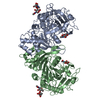
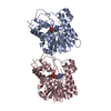
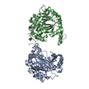
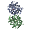
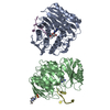
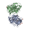
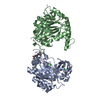
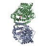
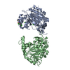
Protein , 1 types, 2 molecules AB
#1: Protein
Lysosomalprotectiveprotein / Carboxypeptidase C / Carboxypeptidase L / Cathepsin A / Protective protein cathepsin A / PPCA / ...Carboxypeptidase C / Carboxypeptidase L / Cathepsin A / Protective protein cathepsin A / PPCA / Protective protein for beta-galactosidase
Mass: 48655.582 Da / Num. of mol.: 2 Source method: isolated from a genetically manipulated source Details: Selected with blasticidin / Source: (gene. exp.) Homo sapiens (human) / Gene: CTSA, PPGB / Plasmid: pIB/V5-His-TOPO TA / Production host: Trichoplusia ni (cabbage looper) / Strain (production host): HI-FIVE / References: UniProt: P10619, carboxypeptidase C
Mass: 18.015 Da / Num. of mol.: 4 / Source method: isolated from a natural source / Formula: H2O
-
Details
Has protein modification
Y
Sequence details
AN UNIDENTIFIED PROTEASE CONVERTED THE ZYMOGEN INTO ACTIVE ENZYME. PUTATIVE TERMINI ASSUME ...AN UNIDENTIFIED PROTEASE CONVERTED THE ZYMOGEN INTO ACTIVE ENZYME. PUTATIVE TERMINI ASSUME PROTEOLYTIC REMOVAL OF 263-292, CONSISTENT WITH MASS SPECTROMETRY AND N-TERMINAL SEQUENCING DATA.
-
Experimental details
-
Experiment
Experiment
Method: X-RAY DIFFRACTION / Number of used crystals: 1
-
Sample preparation
Crystal
Density Matthews: 2.69 Å3/Da / Density % sol: 54.31 %
Crystal grow
Temperature: 293 K / Method: vapor diffusion, sitting drop / pH: 7 Details: 10% PEG 3350, 0.1M sodium formate, pH 7.0, VAPOR DIFFUSION, SITTING DROP, temperature 293K
In the structure databanks used in Yorodumi, some data are registered as the other names, "COVID-19 virus" and "2019-nCoV". Here are the details of the virus and the list of structure data.
Jan 31, 2019. EMDB accession codes are about to change! (news from PDBe EMDB page)
EMDB accession codes are about to change! (news from PDBe EMDB page)
The allocation of 4 digits for EMDB accession codes will soon come to an end. Whilst these codes will remain in use, new EMDB accession codes will include an additional digit and will expand incrementally as the available range of codes is exhausted. The current 4-digit format prefixed with “EMD-” (i.e. EMD-XXXX) will advance to a 5-digit format (i.e. EMD-XXXXX), and so on. It is currently estimated that the 4-digit codes will be depleted around Spring 2019, at which point the 5-digit format will come into force.
The EM Navigator/Yorodumi systems omit the EMD- prefix.
Related info.:Q: What is EMD? / ID/Accession-code notation in Yorodumi/EM Navigator
Yorodumi is a browser for structure data from EMDB, PDB, SASBDB, etc.
This page is also the successor to EM Navigator detail page, and also detail information page/front-end page for Omokage search.
The word "yorodu" (or yorozu) is an old Japanese word meaning "ten thousand". "mi" (miru) is to see.
Related info.:EMDB / PDB / SASBDB / Comparison of 3 databanks / Yorodumi Search / Aug 31, 2016. New EM Navigator & Yorodumi / Yorodumi Papers / Jmol/JSmol / Function and homology information / Changes in new EM Navigator and Yorodumi
 Movie
Movie Controller


 Open data
 Basic information
 Components
 Keywords
 Function and homology information
 Homo sapiens (human)
 X-RAY DIFFRACTION /
  Authors
 Citation
 Structure visualization
 Downloads & links
 Other downloads
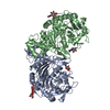

 PDBj
PDBj


 Assembly
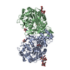
 Trichoplusia ni (cabbage looper) / Strain (production host): HI-FIVE / References: UniProt: P10619, carboxypeptidase C
 Type: D-saccharide, beta linking / Mass: 221.208 Da / Num. of mol.: 2

 Mass: 92.094 Da / Num. of mol.: 2 / Source method: obtained synthetically / Formula: C3H8O3
 Sample preparation
 / Beamline: 24-ID-C / Wavelength: 0.9792 Å
 Processing