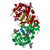
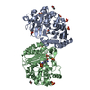
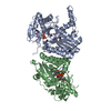
[English] 日本語
Yorodumi
- PDB-4ci9: Crystal structure of cathepsin A, apo-structure -

+
Open data


ID or keywords:

Loading...

-
Basic information

Entry
Database: PDB / ID: 4ci9
TitleCrystal structure of cathepsin A, apo-structure
ComponentsLYSOSOMAL PROTECTIVE PROTEIN
KeywordsHYDROLASE / DRUG DISCOVERY / SERINE CARBOXYPEPTIDASE / CARDIOVASCULAR DRUG / HEART FAILURE / ENDOTHELIN / TETRAHEDRAL INTERMEDIATE / COVALENT INHIBITOR
Function / homology
Function and homology information


carboxypeptidase C / Defective NEU1 causes sialidosis / serine-type carboxypeptidase activity / Sialic acid metabolism / regulation of chaperone-mediated autophagy / Glycosphingolipid catabolism / negative regulation of chaperone-mediated autophagy / carboxypeptidase activity / MHC class II antigen presentation / lysosomal lumen ...carboxypeptidase C / Defective NEU1 causes sialidosis / serine-type carboxypeptidase activity / Sialic acid metabolism / regulation of chaperone-mediated autophagy / Glycosphingolipid catabolism / negative regulation of chaperone-mediated autophagy / carboxypeptidase activity / MHC class II antigen presentation / lysosomal lumen / enzyme activator activity / intracellular protein transport / regulation of protein stability / azurophil granule lumen / lysosome / intracellular membrane-bounded organelle / Neutrophil degranulation / endoplasmic reticulum / proteolysis / extracellular exosome / extracellular region / membrane
Similarity search - Function
Serine carboxypeptidase, serine active site / Serine carboxypeptidases, serine active site. / Peptidase S10, serine carboxypeptidase / Serine carboxypeptidases, histidine active site / Serine carboxypeptidase / Serine carboxypeptidases, histidine active site. / Alpha/Beta hydrolase fold, catalytic domain / Alpha/Beta hydrolase fold / Rossmann fold / 3-Layer(aba) Sandwich / Alpha Beta
Similarity search - Domain/homology
ACETATE ION / Lysosomal protective protein
Similarity search - Component
Biological speciesHOMO SAPIENS (human)
MethodX-RAY DIFFRACTION / SYNCHROTRON / MOLECULAR REPLACEMENT / Resolution: 1.58 Å
AuthorsSchreuder, H.A. / Liesum, A. / Kroll, K. / Boehnisch, B. / Buning, C. / Ruf, S. / Buning, C. / Sadowski, T.
Citation
Journal: Biochem. Biophys. Res. Commun. / Year: 2014
Title: Crystal structure of cathepsin A, a novel target for the treatment of cardiovascular diseases.
Authors: Schreuder, H.A. / Liesum, A. / Kroll, K. / Bohnisch, B. / Buning, C. / Ruf, S. / Sadowski, T.
#1: Journal: J.Med.Chem. / Year: 2012
Title: Novel Beta-Amino Acid Derivatives as Inhibitors of Cathepsin A.
Authors: Ruf, S. / Buning, C. / Schreuder, H. / Horstick, G. / Linz, W. / Olpp, T. / Pernerstorfer, J. / Hiss, K. / Kroll, K. / Kannt, A. / Kohlmann, M. / Linz, D. / Hubschle, T. / Rutten, H. / ...Authors: Ruf, S. / Buning, C. / Schreuder, H. / Horstick, G. / Linz, W. / Olpp, T. / Pernerstorfer, J. / Hiss, K. / Kroll, K. / Kannt, A. / Kohlmann, M. / Linz, D. / Hubschle, T. / Rutten, H. / Wirth, K. / Schmidt, T. / Sadowski, T.
History
DepositionDec 6, 2013Deposition site: PDBE / Processing site: PDBE
Revision 1.0Feb 26, 2014Provider: repository / Type: Initial release
Revision 1.1Apr 2, 2014Group: Database references
Revision 1.2Mar 13, 2019Group: Data collection / Database references ...Data collection / Database references / Derived calculations / Experimental preparation / Other
Category: citation / citation_author ...citation / citation_author / exptl_crystal_grow / pdbx_database_proc / pdbx_database_status / struct_biol / struct_conn
Item: _citation.journal_abbrev / _citation.journal_id_ISSN ..._citation.journal_abbrev / _citation.journal_id_ISSN / _citation.page_last / _citation.pdbx_database_id_DOI / _citation.title / _citation_author.name / _exptl_crystal_grow.method / _exptl_crystal_grow.temp / _pdbx_database_status.recvd_author_approval / _struct_conn.pdbx_leaving_atom_flag
Revision 2.0Jul 29, 2020Group: Advisory / Atomic model ...Advisory / Atomic model / Data collection / Derived calculations / Other / Structure summary
Category: atom_site / chem_comp ...atom_site / chem_comp / entity / pdbx_branch_scheme / pdbx_chem_comp_identifier / pdbx_database_status / pdbx_entity_branch / pdbx_entity_branch_descriptor / pdbx_entity_branch_link / pdbx_entity_branch_list / pdbx_entity_nonpoly / pdbx_nonpoly_scheme / pdbx_struct_assembly_gen / pdbx_struct_special_symmetry / pdbx_unobs_or_zero_occ_atoms / struct_asym / struct_conn / struct_site / struct_site_gen
Item: _atom_site.B_iso_or_equiv / _atom_site.Cartn_x ..._atom_site.B_iso_or_equiv / _atom_site.Cartn_x / _atom_site.Cartn_y / _atom_site.Cartn_z / _atom_site.auth_asym_id / _atom_site.auth_atom_id / _atom_site.auth_comp_id / _atom_site.auth_seq_id / _atom_site.label_asym_id / _atom_site.label_atom_id / _atom_site.label_comp_id / _atom_site.label_entity_id / _atom_site.occupancy / _atom_site.type_symbol / _chem_comp.name / _chem_comp.type / _entity.formula_weight / _entity.pdbx_description / _entity.pdbx_number_of_molecules / _entity.src_method / _entity.type / _pdbx_database_status.status_code_sf / _pdbx_struct_assembly_gen.asym_id_list / _pdbx_struct_special_symmetry.label_asym_id / _pdbx_unobs_or_zero_occ_atoms.label_asym_id / _struct_conn.pdbx_role / _struct_conn.ptnr1_auth_asym_id / _struct_conn.ptnr1_auth_seq_id / _struct_conn.ptnr1_label_asym_id / _struct_conn.ptnr2_auth_asym_id / _struct_conn.ptnr2_auth_seq_id / _struct_conn.ptnr2_label_asym_id
Description: Carbohydrate remediation / Provider: repository / Type: Remediation
Revision 2.1Dec 20, 2023Group: Data collection / Database references ...Data collection / Database references / Refinement description / Structure summary
Category: chem_comp / chem_comp_atom ...chem_comp / chem_comp_atom / chem_comp_bond / database_2 / pdbx_initial_refinement_model
Item: _chem_comp.pdbx_synonyms / _database_2.pdbx_DOI / _database_2.pdbx_database_accession
Revision 2.2Nov 20, 2024Group: Structure summary / Category: pdbx_entry_details / pdbx_modification_feature / Item: _pdbx_entry_details.has_protein_modification

-
Structure visualization

Structure viewerMolecule:
MolmilJmol/JSmol

Downloads & links

-
Assembly

Deposited unit
A: LYSOSOMAL PROTECTIVE PROTEIN
hetero molecules


Theoretical massNumber of molelcules
Total (without water)53,1559
Polymers51,8291
Non-polymers1,3258
Water11,061614
1
A: LYSOSOMAL PROTECTIVE PROTEIN
hetero molecules

A: LYSOSOMAL PROTECTIVE PROTEIN
hetero molecules


Theoretical massNumber of molelcules
Total (without water)106,30918
Polymers103,6592
Non-polymers2,65116
Water362
TypeNameSymmetry operationNumber
identity operation1_555x,y,z1
crystal symmetry operation2_455-x-1,y,-z1
Buried area8430 Å2
ΔGint-28.5 kcal/mol
Surface area33550 Å2
MethodPISA
Unit cell
Length a, b, c (Å)90.310, 102.030, 48.550
Angle α, β, γ (deg.)90.00, 101.27, 90.00
Int Tables number5
Space group name H-MC121
Components on special symmetry positions
IDModelComponents
11A-2130-

HOH

21A-2350-

HOH

31A-2385-

HOH

-
Components

-
Protein / Sugars , 2 types, 3 molecules A

#1: Protein LYSOSOMAL PROTECTIVE PROTEIN / CARBOXYPEPTIDASE C / CARBOXYPEPTIDASE L / CATHEPSIN A / PROTECTIVE PROTEIN CATHEPSIN A / PPCA / ...CARBOXYPEPTIDASE C / CARBOXYPEPTIDASE L / CATHEPSIN A / PROTECTIVE PROTEIN CATHEPSIN A / PPCA / PROTECTIVE PROTEIN FOR BETA-GALACTOSIDASE


Mass: 51829.359 Da / Num. of mol.: 1
Source method: isolated from a genetically manipulated source
Details: ACTIVATED WITH TRYPSIN-SEPHAROSE / Source: (gene. exp.) HOMO SAPIENS (human) / Cell line (production host): SF9 / Production host: SPODOPTERA FRUGIPERDA (fall armyworm) / References: UniProt: P10619, carboxypeptidase C
#2: Polysaccharide 2-acetamido-2-deoxy-beta-D-glucopyranose-(1-4)-2-acetamido-2-deoxy-beta-D-glucopyranose


Type: oligosaccharide / Mass: 424.401 Da / Num. of mol.: 2
Source method: isolated from a genetically manipulated source
DescriptorTypeProgram
DGlcpNAcb1-4DGlcpNAcb1-Glycam Condensed SequenceGMML 1.0
WURCS=2.0/1,2,1/[a2122h-1b_1-5_2*NCC/3=O]/1-1/a4-b1WURCSPDB2Glycan 1.1.0
[]{[(4+1)][b-D-GlcpNAc]{[(4+1)][b-D-GlcpNAc]{}}}LINUCSPDB-CARE

-
Non-polymers , 5 types, 620 molecules

#3: Chemical ChemComp-DMS / DIMETHYL SULFOXIDE


Mass: 78.133 Da / Num. of mol.: 1 / Source method: obtained synthetically / Formula: C2H6OS / Comment: DMSO, precipitant*YM
#4: Chemical ChemComp-ACT / ACETATE ION


Mass: 59.044 Da / Num. of mol.: 2 / Source method: obtained synthetically / Formula: C2H3O2
#5: Chemical ChemComp-GOL / GLYCEROL / GLYCERIN / PROPANE-1,2,3-TRIOL


Mass: 92.094 Da / Num. of mol.: 2 / Source method: obtained synthetically / Formula: C3H8O3
#6: Chemical ChemComp-SO4 / SULFATE ION


Mass: 96.063 Da / Num. of mol.: 1 / Source method: obtained synthetically / Formula: SO4
#7: Water ChemComp-HOH / water


Mass: 18.015 Da / Num. of mol.: 614 / Source method: isolated from a natural source / Formula: H2O

-
Details

Has protein modificationY
Sequence detailsAT THE C-TERMINUS, ONE EXTRA GLU IS PRESENT AS A LEFTOVER FROM A MYC-TAG

-
Experimental details

-
Experiment

ExperimentMethod: X-RAY DIFFRACTION / Number of used crystals: 1

-
Sample preparation

CrystalDensity Matthews: 2.34 Å3/Da / Density % sol: 47.4 % / Description: NONE
Crystal growTemperature: 277 K / Method: vapor diffusion, hanging drop / pH: 4.6
Details: CATHEPSIN A WAS CRYSTALLIZED USING THE HANGING DROP METHOD: 1 UL OF PROTEIN SOLUTION, CONTAINING 6.5 MG/ML CATHEPSIN A, 25 MM TRIS-HCL (PH 8.0) AND 300 MM NACL, WAS MIXED WITH 1 UL RESERVOIR ...Details: CATHEPSIN A WAS CRYSTALLIZED USING THE HANGING DROP METHOD: 1 UL OF PROTEIN SOLUTION, CONTAINING 6.5 MG/ML CATHEPSIN A, 25 MM TRIS-HCL (PH 8.0) AND 300 MM NACL, WAS MIXED WITH 1 UL RESERVOIR SOLUTION, CONTAINING 100 MM NAACETATE (PH 4.5), 18-20% PEG400 AND 100 MM CDCL2, AND SET TO EQUILIBRATE AT 4DEG.C. ROD-SHAPED CRYSTALS APPEARED IN ABOUT ONE WEEK.

-
Data collection

DiffractionMean temperature: 100 K
Diffraction sourceSource: SYNCHROTRON / Site: SLS / Beamline: X10SA / Wavelength: 1
DetectorType: DECTRIS PILATUS / Detector: PIXEL / Date: Mar 12, 2012
RadiationProtocol: SINGLE WAVELENGTH / Monochromatic (M) / Laue (L): M / Scattering type: x-ray
Radiation wavelengthWavelength: 1 Å / Relative weight: 1
ReflectionResolution: 1.58→33.44 Å / Num. obs: 56973 / % possible obs: 96.6 % / Observed criterion σ(I): -3 / Redundancy: 3.3 % / Biso Wilson estimate: 19.14 Å2 / Rmerge(I) obs: 0.04 / Net I/σ(I): 18.1
Reflection shellResolution: 1.58→1.66 Å / Redundancy: 2.3 % / Rmerge(I) obs: 0.35 / Mean I/σ(I) obs: 2.3 / % possible all: 94.3

-
Processing

Software
NameVersionClassification
BUSTER2.11.2refinement
XDSdata reduction
SCALAdata scaling
PHASERphasing
RefinementMethod to determine structure: MOLECULAR REPLACEMENT
Starting model: PDB ENTRY 4AZ0
Resolution: 1.58→33.44 Å / Cor.coef. Fo:Fc: 0.9584 / Cor.coef. Fo:Fc free: 0.9471 / SU R Cruickshank DPI: 0.083 / Cross valid method: THROUGHOUT / σ(F): 0 / SU R Blow DPI: 0.089 / SU Rfree Blow DPI: 0.085 / SU Rfree Cruickshank DPI: 0.081
RfactorNum. reflection% reflectionSelection details
Rfree0.1905 2843 5 %RANDOM
Rwork0.1649 ---
obs0.1662 56890 96.52 %-
Displacement parametersBiso mean: 23.01 Å2
Baniso -1Baniso -2Baniso -3
1-3.1215 Å20 Å22.4778 Å2
2--0.3008 Å20 Å2
3----3.4223 Å2
Refine analyzeLuzzati coordinate error obs: 0.165 Å
Refinement stepCycle: LAST / Resolution: 1.58→33.44 Å
ProteinNucleic acidLigandSolventTotal
Num. atoms3302 0 84 614 4000
Refine LS restraints
Refine-IDTypeDev idealNumberRestraint functionWeight
X-RAY DIFFRACTIONt_bond_d0.0073576HARMONIC2
X-RAY DIFFRACTIONt_angle_deg0.914882HARMONIC2
X-RAY DIFFRACTIONt_dihedral_angle_d1213SINUSOIDAL2
X-RAY DIFFRACTIONt_incorr_chiral_ct
X-RAY DIFFRACTIONt_pseud_angle
X-RAY DIFFRACTIONt_trig_c_planes99HARMONIC2
X-RAY DIFFRACTIONt_gen_planes520HARMONIC5
X-RAY DIFFRACTIONt_it3576HARMONIC20
X-RAY DIFFRACTIONt_nbd
X-RAY DIFFRACTIONt_omega_torsion2.84
X-RAY DIFFRACTIONt_other_torsion16.59
X-RAY DIFFRACTIONt_improper_torsion
X-RAY DIFFRACTIONt_chiral_improper_torsion441SEMIHARMONIC5
X-RAY DIFFRACTIONt_sum_occupancies12HARMONIC1
X-RAY DIFFRACTIONt_utility_distance
X-RAY DIFFRACTIONt_utility_angle
X-RAY DIFFRACTIONt_utility_torsion
X-RAY DIFFRACTIONt_ideal_dist_contact4641SEMIHARMONIC4
LS refinement shellResolution: 1.58→1.62 Å / Total num. of bins used: 20
RfactorNum. reflection% reflection
Rfree0.234 202 5.07 %
Rwork0.2196 3780 -
all0.2204 3982 -
obs--96.52 %

+
About Yorodumi

-
News

-
Feb 9, 2022. New format data for meta-information of EMDB entries

New format data for meta-information of EMDB entries

  • Version 3 of the EMDB header file is now the official format.
  • The previous official version 1.9 will be removed from the archive.

Related info.:EMDB header

External links:wwPDB to switch to version 3 of the EMDB data model

-
Aug 12, 2020. Covid-19 info

Covid-19 info

URL: https://pdbj.org/emnavi/covid19.php

New page: Covid-19 featured information page in EM Navigator.

Related info.:Covid-19 info / Mar 5, 2020. Novel coronavirus structure data

+
Mar 5, 2020. Novel coronavirus structure data

Novel coronavirus structure data

Related info.:Yorodumi Speices / Aug 12, 2020. Covid-19 info

External links:COVID-19 featured content - PDBj / Molecule of the Month (242):Coronavirus Proteases

+
Jan 31, 2019. EMDB accession codes are about to change! (news from PDBe EMDB page)

EMDB accession codes are about to change! (news from PDBe EMDB page)

  • The allocation of 4 digits for EMDB accession codes will soon come to an end. Whilst these codes will remain in use, new EMDB accession codes will include an additional digit and will expand incrementally as the available range of codes is exhausted. The current 4-digit format prefixed with “EMD-” (i.e. EMD-XXXX) will advance to a 5-digit format (i.e. EMD-XXXXX), and so on. It is currently estimated that the 4-digit codes will be depleted around Spring 2019, at which point the 5-digit format will come into force.
  • The EM Navigator/Yorodumi systems omit the EMD- prefix.

Related info.:Q: What is EMD? / ID/Accession-code notation in Yorodumi/EM Navigator

External links:EMDB Accession Codes are Changing Soon! / Contact to PDBj

+
Jul 12, 2017. Major update of PDB

Major update of PDB

  • wwPDB released updated PDB data conforming to the new PDBx/mmCIF dictionary.
  • This is a major update changing the version number from 4 to 5, and with Remediation, in which all the entries are updated.
  • In this update, many items about electron microscopy experimental information are reorganized (e.g. em_software).
  • Now, EM Navigator and Yorodumi are based on the updated data.

External links:wwPDB Remediation / Enriched Model Files Conforming to OneDep Data Standards Now Available in the PDB FTP Archive

-
Yorodumi

Thousand views of thousand structures

  • Yorodumi is a browser for structure data from EMDB, PDB, SASBDB, etc.
  • This page is also the successor to EM Navigator detail page, and also detail information page/front-end page for Omokage search.
  • The word "yorodu" (or yorozu) is an old Japanese word meaning "ten thousand". "mi" (miru) is to see.

Related info.:EMDB / PDB / SASBDB / Comparison of 3 databanks / Yorodumi Search / Aug 31, 2016. New EM Navigator & Yorodumi / Yorodumi Papers / Jmol/JSmol / Function and homology information / Changes in new EM Navigator and Yorodumi

Read more