 Movie
Movie Controller


+ Open data

- Basic information
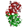
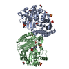
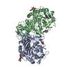
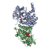
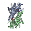
| Entry | Database: PDB / ID: 4cia | ||||||
|---|---|---|---|---|---|---|---|
| Title | Crystal structure of cathepsin A, complexed with compound 1 | ||||||
 Components Components | LYSOSOMAL PROTECTIVE PROTEIN | ||||||
 Keywords Keywords | HYDROLASE / SERINE CARBOXYPEPTIDASE / CARDIOVASCULAR DRUG / HEART FAILURE / ENDOTHELIN / TETRAHEDRAL INTERMEDIATE / COVALENT INHIBITOR | ||||||
| Function / homology |  Function and homology information Function and homology informationcarboxypeptidase C / Defective NEU1 causes sialidosis / serine-type carboxypeptidase activity / Sialic acid metabolism / regulation of chaperone-mediated autophagy / Glycosphingolipid catabolism / negative regulation of chaperone-mediated autophagy / carboxypeptidase activity / MHC class II antigen presentation / lysosomal lumen ...carboxypeptidase C / Defective NEU1 causes sialidosis / serine-type carboxypeptidase activity / Sialic acid metabolism / regulation of chaperone-mediated autophagy / Glycosphingolipid catabolism / negative regulation of chaperone-mediated autophagy / carboxypeptidase activity / MHC class II antigen presentation / lysosomal lumen / regulation of protein stability / intracellular protein transport / enzyme activator activity / azurophil granule lumen / lysosome / Neutrophil degranulation / endoplasmic reticulum / proteolysis / extracellular exosome / extracellular region / membrane Similarity search - Function | ||||||
| Biological species |  HOMO SAPIENS (human) HOMO SAPIENS (human) | ||||||
| Method |  X-RAY DIFFRACTION / SYNCHROTRON / MOLECULAR REPLACEMENT / Resolution: 1.98 Å X-RAY DIFFRACTION / SYNCHROTRON / MOLECULAR REPLACEMENT / Resolution: 1.98 Å | ||||||
 Authors Authors | Schreuder, H.A. / Liesum, A. / Kroll, K. / Boehnisch, B. / Buning, C. / Ruf, S. / Buning, C. / Sadowski, T. | ||||||
 Citation Citation | Journal: Biochem. Biophys. Res. Commun. / Year: 2014 Title: Crystal structure of cathepsin A, a novel target for the treatment of cardiovascular diseases. Authors: Schreuder, H.A. / Liesum, A. / Kroll, K. / Bohnisch, B. / Buning, C. / Ruf, S. / Sadowski, T. #1: Journal: J.Med.Chem. / Year: 2012Title: Novel Beta-Amino Acid Derivatives as Inhibitors of Cathepsin A. Authors: Ruf, S. / Buning, C. / Schreuder, H. / Horstick, G. / Linz, W. / Olpp, T. / Pernerstorfer, J. / Hiss, K. / Kroll, K. / Kannt, A. / Kohlmann, M. / Linz, D. / Hubschle, T. / Rutten, H. / ...Authors: Ruf, S. / Buning, C. / Schreuder, H. / Horstick, G. / Linz, W. / Olpp, T. / Pernerstorfer, J. / Hiss, K. / Kroll, K. / Kannt, A. / Kohlmann, M. / Linz, D. / Hubschle, T. / Rutten, H. / Wirth, K. / Schmidt, T. / Sadowski, T. | ||||||
| History |
|
- Structure visualization
| Structure viewer | Molecule: MolmilJmol/JSmol |
|---|
- Downloads & links
-Download
| PDBx/mmCIF format | 4cia.cif.gz | 109.6 KB | Display | PDBx/mmCIF format |
|---|---|---|---|---|
| PDB format | pdb4cia.ent.gz | 81.7 KB | Display | PDB format |
| PDBx/mmJSON format | 4cia.json.gz | Tree view | PDBx/mmJSON format | |
| Others |  Other downloads Other downloads |
-Validation report
| Arichive directory | https://data.pdbj.org/pub/pdb/validation_reports/ci/4ciaftp://data.pdbj.org/pub/pdb/validation_reports/ci/4cia | HTTPS FTP |
|---|
-Related structure data
| Related structure data | 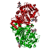 4ci9C 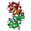 4cibC  4az0S C: citing same article ( S: Starting model for refinement |
|---|---|
| Similar structure data |


-Links
 PDBj
PDBj


- Assembly
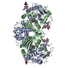
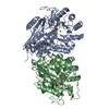
| Deposited unit | 
| |||||||||
|---|---|---|---|---|---|---|---|---|---|---|
| 1 | 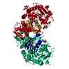
| |||||||||
| Unit cell |
| |||||||||
| Components on special symmetry positions |
|
-Components
| #1: Protein | Mass: 51829.359 Da / Num. of mol.: 1 Source method: isolated from a genetically manipulated source Details: ACTIVATED WITH TRYPSIN-SEPHAROSE / Source: (gene. exp.) HOMO SAPIENS (human) / Cell line (production host): SF9 / Production host:   SPODOPTERA FRUGIPERDA (fall armyworm) / References: UniProt: P10619, carboxypeptidase C SPODOPTERA FRUGIPERDA (fall armyworm) / References: UniProt: P10619, carboxypeptidase C | ||||||||
|---|---|---|---|---|---|---|---|---|---|
| #2: Chemical | ChemComp-6KZ /   Mass: 459.539 Da / Num. of mol.: 1 / Source method: obtained synthetically / Formula: C23H33N5O5 Mass: 459.539 Da / Num. of mol.: 1 / Source method: obtained synthetically / Formula: C23H33N5O5 | ||||||||
| #3: Chemical | ChemComp-CD /   Mass: 112.411 Da / Num. of mol.: 7 / Source method: obtained synthetically / Formula: Cd Mass: 112.411 Da / Num. of mol.: 7 / Source method: obtained synthetically / Formula: Cd#4: Sugar |   Type: D-saccharide, beta linking / Mass: 221.208 Da / Num. of mol.: 2 Type: D-saccharide, beta linking / Mass: 221.208 Da / Num. of mol.: 2Source method: isolated from a genetically manipulated source Formula: C8H15NO6 #5: Water | ChemComp-HOH / |  Mass: 18.015 Da / Num. of mol.: 358 / Source method: isolated from a natural source / Formula: H2O Mass: 18.015 Da / Num. of mol.: 358 / Source method: isolated from a natural source / Formula: H2OHas protein modification | Y | Sequence details | AT THE N-TERMINUS, 2 RESIDUES FROM PREPROMELLITIN SIGNAL SEQUENCE ARE PRESENT, AT THE C-TERMINUS, 1 ...AT THE N-TERMINUS, 2 RESIDUES FROM PREPROMELL | |
-Experimental details
-Experiment
| Experiment | Method: X-RAY DIFFRACTION / Number of used crystals: 1 |
|---|
- Sample preparation
| Crystal | Density Matthews: 2.3 Å3/Da / Density % sol: 46.5 % / Description: NONE |
|---|---|
| Crystal grow | Temperature: 277 K / Method: vapor diffusion, hanging drop / pH: 4.6 Details: CATHEPSIN A WAS CRYSTALLIZED USING THE HANGING DROP METHOD: 1 UL OF PROTEIN SOLUTION, CONTAINING 6.5 MG/ML CATHEPSIN A, 25 MM TRIS-HCL (PH 8.0) AND 300 MM NACL, WAS MIXED WITH 1 UL RESERVOIR ...Details: CATHEPSIN A WAS CRYSTALLIZED USING THE HANGING DROP METHOD: 1 UL OF PROTEIN SOLUTION, CONTAINING 6.5 MG/ML CATHEPSIN A, 25 MM TRIS-HCL (PH 8.0) AND 300 MM NACL, WAS MIXED WITH 1 UL RESERVOIR SOLUTION, CONTAINING 100 MM NAACETATE (PH 4.5), 18-20% PEG400 AND 100 MM CDCL2, AND SET TO EQUILIBRATE AT 4DEG.C. ROD-SHAPED CRYSTALS APPEARED IN ABOUT ONE WEEK. |
-Data collection
| Diffraction | Mean temperature: 100 K |
|---|---|
| Diffraction source | Source: SYNCHROTRON / Site: ESRF  / Beamline: ID14-4 / Wavelength: 0.9395 / Beamline: ID14-4 / Wavelength: 0.9395 |
| Detector | Type: ADSC CCD / Detector: CCD / Date: Sep 11, 2007 |
| Radiation | Protocol: SINGLE WAVELENGTH / Monochromatic (M) / Laue (L): M / Scattering type: x-ray |
| Radiation wavelength | Wavelength: 0.9395 Å / Relative weight: 1 |
| Reflection | Resolution: 1.97→66.25 Å / Num. obs: 28737 / % possible obs: 96.2 % / Observed criterion σ(I): -3 / Redundancy: 3.7 % / Rmerge(I) obs: 0.08 / Net I/σ(I): 11.47 |
| Reflection shell | Resolution: 1.97→2.07 Å / Redundancy: 3.3 % / Rmerge(I) obs: 0.28 / Mean I/σ(I) obs: 4.12 / % possible all: 73.5 |
- Processing
| Software |
| ||||||||||||||||||||||||||||||||||||||||||||||||||||||||||||||||||||||||||||||||||||||||||||||||||||||||||||||||||||||||||||||||||||||||||||||||||||||||||||||||||||||||||||||||||||||
|---|---|---|---|---|---|---|---|---|---|---|---|---|---|---|---|---|---|---|---|---|---|---|---|---|---|---|---|---|---|---|---|---|---|---|---|---|---|---|---|---|---|---|---|---|---|---|---|---|---|---|---|---|---|---|---|---|---|---|---|---|---|---|---|---|---|---|---|---|---|---|---|---|---|---|---|---|---|---|---|---|---|---|---|---|---|---|---|---|---|---|---|---|---|---|---|---|---|---|---|---|---|---|---|---|---|---|---|---|---|---|---|---|---|---|---|---|---|---|---|---|---|---|---|---|---|---|---|---|---|---|---|---|---|---|---|---|---|---|---|---|---|---|---|---|---|---|---|---|---|---|---|---|---|---|---|---|---|---|---|---|---|---|---|---|---|---|---|---|---|---|---|---|---|---|---|---|---|---|---|---|---|---|---|
| Refinement | Method to determine structure: MOLECULAR REPLACEMENT Starting model: PDB ENTRY 4AZ0 Resolution: 1.98→36.11 Å / Cor.coef. Fo:Fc: 0.95 / Cor.coef. Fo:Fc free: 0.905 / SU B: 4.15 / SU ML: 0.115 / Cross valid method: THROUGHOUT / ESU R: 0.188 / ESU R Free: 0.174 / Stereochemistry target values: MAXIMUM LIKELIHOOD / Details: HYDROGENS HAVE BEEN USED IF PRESENT IN THE INPUT
| ||||||||||||||||||||||||||||||||||||||||||||||||||||||||||||||||||||||||||||||||||||||||||||||||||||||||||||||||||||||||||||||||||||||||||||||||||||||||||||||||||||||||||||||||||||||
| Solvent computation | Ion probe radii: 0.8 Å / Shrinkage radii: 0.8 Å / VDW probe radii: 1.4 Å / Solvent model: MASK | ||||||||||||||||||||||||||||||||||||||||||||||||||||||||||||||||||||||||||||||||||||||||||||||||||||||||||||||||||||||||||||||||||||||||||||||||||||||||||||||||||||||||||||||||||||||
| Displacement parameters | Biso mean: 20.508 Å2
| ||||||||||||||||||||||||||||||||||||||||||||||||||||||||||||||||||||||||||||||||||||||||||||||||||||||||||||||||||||||||||||||||||||||||||||||||||||||||||||||||||||||||||||||||||||||
| Refinement step | Cycle: LAST / Resolution: 1.98→36.11 Å
| ||||||||||||||||||||||||||||||||||||||||||||||||||||||||||||||||||||||||||||||||||||||||||||||||||||||||||||||||||||||||||||||||||||||||||||||||||||||||||||||||||||||||||||||||||||||
| Refine LS restraints |
|