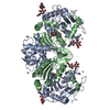
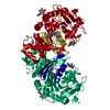
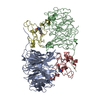
Lysosomalprotectiveprotein / Carboxypeptidase C / Carboxypeptidase L / Cathepsin A / Protective protein cathepsin A / PPCA / ...Carboxypeptidase C / Carboxypeptidase L / Cathepsin A / Protective protein cathepsin A / PPCA / Protective protein for beta-galactosidase
Mass: 52406.906 Da / Num. of mol.: 1 Source method: isolated from a genetically manipulated source Details: cell line / Source: (gene. exp.) Homo sapiens (human) / Gene: CTSA, PPGB / Plasmid: pcDNA3.4 / Cell line (production host): Expi293 / Production host: Homo sapiens (human) / References: UniProt: P10619, carboxypeptidase C
Mass: 18.015 Da / Num. of mol.: 173 / Source method: isolated from a natural source / Formula: H2O
-
Details
Has ligand of interest
Y
-
Experimental details
-
Experiment
Experiment
Method: X-RAY DIFFRACTION / Number of used crystals: 1
-
Sample preparation
Crystal
Density Matthews: 2.09 Å3/Da / Density % sol: 41.23 %
Crystal grow
Temperature: 277 K / Method: vapor diffusion, hanging drop / pH: 4.6 Details: Crystallized in Crystal Screen Cryo II condition #12 (0.095 M cadmium chloride hydrate, 0.095 M sodium acetate trihydrate pH 4.6, 28.5% v/v PEG 400, 5% glycerol). Mounted in 30% glycerol and ...Details: Crystallized in Crystal Screen Cryo II condition #12 (0.095 M cadmium chloride hydrate, 0.095 M sodium acetate trihydrate pH 4.6, 28.5% v/v PEG 400, 5% glycerol). Mounted in 30% glycerol and 70% CS Cryo II condition #12. Protein was inhibited with DFP in 25 mM Tris pH 8, 300 mM NaCl at R.T.
-
Data collection
Diffraction
Mean temperature: 100 K / Serial crystal experiment: N
Diffraction source
Source: SEALED TUBE / Type: BRUKER D8 QUEST / Wavelength: 1.54 Å
Detector
Type: Bruker PHOTON III / Detector: PIXEL / Date: Nov 9, 2018
Radiation
Protocol: SINGLE WAVELENGTH / Monochromatic (M) / Laue (L): M / Scattering type: x-ray
Radiation wavelength
Wavelength: 1.54 Å / Relative weight: 1
Reflection
Resolution: 2.21→41.7 Å / Num. obs: 21594 / % possible obs: 99.8 % / Redundancy: 5.12 % / Rmerge(I) obs: 0.0836 / Net I/σ(I): 11.55
Reflection shell
Resolution: 2.21→2.31 Å / Redundancy: 5.05 % / Rmerge(I) obs: 0.3905 / Mean I/σ(I) obs: 3.36 / Num. unique obs: 2654 / % possible all: 99.8
-
Processing
Software
Name
Version
Classification
REFMAC
5.8.0258
refinement
PDB_EXTRACT
3.25
dataextraction
APEX
datareduction
APEX
datascaling
PHASER
phasing
Refinement
Method to determine structure: MOLECULAR REPLACEMENT Starting model: 4az3
In the structure databanks used in Yorodumi, some data are registered as the other names, "COVID-19 virus" and "2019-nCoV". Here are the details of the virus and the list of structure data.
Jan 31, 2019. EMDB accession codes are about to change! (news from PDBe EMDB page)
EMDB accession codes are about to change! (news from PDBe EMDB page)
The allocation of 4 digits for EMDB accession codes will soon come to an end. Whilst these codes will remain in use, new EMDB accession codes will include an additional digit and will expand incrementally as the available range of codes is exhausted. The current 4-digit format prefixed with “EMD-” (i.e. EMD-XXXX) will advance to a 5-digit format (i.e. EMD-XXXXX), and so on. It is currently estimated that the 4-digit codes will be depleted around Spring 2019, at which point the 5-digit format will come into force.
The EM Navigator/Yorodumi systems omit the EMD- prefix.
Related info.:Q: What is EMD? / ID/Accession-code notation in Yorodumi/EM Navigator
Yorodumi is a browser for structure data from EMDB, PDB, SASBDB, etc.
This page is also the successor to EM Navigator detail page, and also detail information page/front-end page for Omokage search.
The word "yorodu" (or yorozu) is an old Japanese word meaning "ten thousand". "mi" (miru) is to see.
Related info.:EMDB / PDB / SASBDB / Comparison of 3 databanks / Yorodumi Search / Aug 31, 2016. New EM Navigator & Yorodumi / Yorodumi Papers / Jmol/JSmol / Function and homology information / Changes in new EM Navigator and Yorodumi
 Movie
Movie Controller

 Yorodumi
 Open data
 Basic information
 Components
 Keywords
 Function and homology information
 Homo sapiens (human)
 X-RAY DIFFRACTION /
  Authors
 United States, 1items
  Citation
 Structure visualization
 Downloads & links
 Other downloads
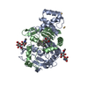


 PDBj
PDBj


 Assembly


 Mass: 59.044 Da / Num. of mol.: 5 / Source method: obtained synthetically / Formula: C2H3O2
 Mass: 112.411 Da / Num. of mol.: 6 / Source method: obtained synthetically / Formula: Cd
 Mass: 92.094 Da / Num. of mol.: 1 / Source method: obtained synthetically / Formula: C3H8O3
 Sample preparation
 Processing