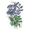
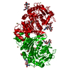
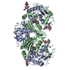
LYSOSOMAL PROTECTIVE PROTEIN ... , 2 types, 2 molecules AB
#1: Protein
LYSOSOMALPROTECTIVEPROTEIN32KDACHAIN / CARBOXYPEPTIDASE C / CARBOXYPEPTIDASE L / CATHEPSIN A / PROTECTIVE PROTEIN CATHEPSIN A / PPCA / ...CARBOXYPEPTIDASE C / CARBOXYPEPTIDASE L / CATHEPSIN A / PROTECTIVE PROTEIN CATHEPSIN A / PPCA / PROTECTIVE PROTEIN FOR BETA-GALACTOSIDASE
Mass: 33773.734 Da / Num. of mol.: 1 Source method: isolated from a genetically manipulated source Details: ACTIVATED WITH TRYPSIN-SEPHAROSE / Source: (gene. exp.) HOMO SAPIENS (human) / Cell line (production host): SF9 / Production host: SPODOPTERA FRUGIPERDA (fall armyworm) / References: UniProt: P10619, carboxypeptidase C
#2: Protein
LYSOSOMALPROTECTIVEPROTEIN20KDACHAIN / CARBOXYPEPTIDASE C / CARBOXYPEPTIDASE L / CATHEPSIN A / PROTECTIVE PROTEIN CATHEPSIN A / PPCA / ...CARBOXYPEPTIDASE C / CARBOXYPEPTIDASE L / CATHEPSIN A / PROTECTIVE PROTEIN CATHEPSIN A / PPCA / PROTECTIVE PROTEIN FOR BETA-GALACTOSIDASE
Mass: 18073.738 Da / Num. of mol.: 1 Source method: isolated from a genetically manipulated source Details: ACTIVATED WITH TRYPSIN-SEPHAROSE / Source: (gene. exp.) HOMO SAPIENS (human) / Cell line (production host): SF9 / Production host: SPODOPTERA FRUGIPERDA (fall armyworm) / References: UniProt: P10619, carboxypeptidase C
Mass: 18.015 Da / Num. of mol.: 308 / Source method: isolated from a natural source / Formula: H2O
-
Details
Has protein modification
Y
Sequence details
AT THE C-TERMINUS, ONE EXTRA GLU IS PRESENT AS A LEFTOVER
-
Experimental details
-
Experiment
Experiment
Method: X-RAY DIFFRACTION / Number of used crystals: 1
-
Sample preparation
Crystal
Density Matthews: 2.3 Å3/Da / Density % sol: 46.6 % / Description: NONE
Crystal grow
Temperature: 277 K / Method: vapor diffusion, hanging drop / pH: 4.6 Details: CATA WAS CRYSTALLIZED USING THE HANGING DROP METHOD: 1 UL OF PROTEIN SOLUTION, CONTAINING 6.5 MG/ML CATHEPSIN A, 25 MM TRIS-HCL (PH 8.0) AND 300 MM NACL, WAS MIXED WITH 1 UL RESERVOIR ...Details: CATA WAS CRYSTALLIZED USING THE HANGING DROP METHOD: 1 UL OF PROTEIN SOLUTION, CONTAINING 6.5 MG/ML CATHEPSIN A, 25 MM TRIS-HCL (PH 8.0) AND 300 MM NACL, WAS MIXED WITH 1 UL RESERVOIR SOLUTION, CONTAINING 100 MM NAACETATE (PH 4.5), 18-20% PEG400 AND 100 MM CDCL2, AND SET TO EQUILIBRATE AT 4 DEG. C. ROD-SHAPED CRYSTALS APPEARED IN ABOUT ONE WEEK.
Resolution: 2.17→44.28 Å / Cor.coef. Fo:Fc: 0.9494 / Cor.coef. Fo:Fc free: 0.9267 / SU R Cruickshank DPI: 0.27 / Cross valid method: THROUGHOUT / σ(F): 0 / SU R Blow DPI: 0.284 / SU Rfree Blow DPI: 0.194 / SU Rfree Cruickshank DPI: 0.193 Details: IDEAL-DIST CONTACT TERM CONTACT SETUP. RESIDUE TYPES WITHOUT CCP4 ATOM TYPE IN LIBRARY=CD. NUMBER OF ATOMS WITH PROPER CCP4 ATOM TYPE=3670. NUMBER WITH APPROX DEFAULT CCP4 ATOM TYPE=0. ...Details: IDEAL-DIST CONTACT TERM CONTACT SETUP. RESIDUE TYPES WITHOUT CCP4 ATOM TYPE IN LIBRARY=CD. NUMBER OF ATOMS WITH PROPER CCP4 ATOM TYPE=3670. NUMBER WITH APPROX DEFAULT CCP4 ATOM TYPE=0. NUMBER TREATED BY BAD NON-BONDED CONTACTS=6.
Num. reflection
% reflection
Selection details
Rfree
1106
4.98 %
RANDOM
obs
22193
98.22 %
-
Displacement parameters
Biso mean: 37.98 Å2
Baniso -1
Baniso -2
Baniso -3
1-
-1.0598 Å2
0 Å2
0.0409 Å2
2-
-
-1.1108 Å2
0 Å2
3-
-
-
2.1705 Å2
Refine analyze
Luzzati coordinate error obs: 0.235 Å
Refinement step
Cycle: LAST / Resolution: 2.17→44.28 Å
Protein
Nucleic acid
Ligand
Solvent
Total
Num. atoms
3278
0
90
308
3676
Refine LS restraints
Refine-ID
Type
Dev ideal
Number
Restraint function
Weight
X-RAY DIFFRACTION
t_bond_d
0.007
3463
HARMONIC
2
X-RAY DIFFRACTION
t_angle_deg
0.95
4721
HARMONIC
2
X-RAY DIFFRACTION
t_dihedral_angle_d
1158
SINUSOIDAL
2
X-RAY DIFFRACTION
t_incorr_chiral_ct
X-RAY DIFFRACTION
t_pseud_angle
X-RAY DIFFRACTION
t_trig_c_planes
94
HARMONIC
2
X-RAY DIFFRACTION
t_gen_planes
518
HARMONIC
5
X-RAY DIFFRACTION
t_it
3463
HARMONIC
20
X-RAY DIFFRACTION
t_nbd
0
SEMIHARMONIC
5
X-RAY DIFFRACTION
t_omega_torsion
2.36
X-RAY DIFFRACTION
t_other_torsion
16.88
X-RAY DIFFRACTION
t_improper_torsion
X-RAY DIFFRACTION
t_chiral_improper_torsion
429
SEMIHARMONIC
5
X-RAY DIFFRACTION
t_sum_occupancies
X-RAY DIFFRACTION
t_utility_distance
X-RAY DIFFRACTION
t_utility_angle
X-RAY DIFFRACTION
t_utility_torsion
X-RAY DIFFRACTION
t_ideal_dist_contact
4190
SEMIHARMONIC
4
LS refinement shell
Resolution: 2.17→2.28 Å / Total num. of bins used: 11
Rfactor
Num. reflection
% reflection
Rfree
0.2892
147
5.44 %
Rwork
0.2299
2554
-
all
0.233
2701
-
obs
-
-
98.22 %
+
About Yorodumi
-
News
-
Feb 9, 2022. New format data for meta-information of EMDB entries
New format data for meta-information of EMDB entries
Version 3 of the EMDB header file is now the official format.
The previous official version 1.9 will be removed from the archive.
In the structure databanks used in Yorodumi, some data are registered as the other names, "COVID-19 virus" and "2019-nCoV". Here are the details of the virus and the list of structure data.
Jan 31, 2019. EMDB accession codes are about to change! (news from PDBe EMDB page)
EMDB accession codes are about to change! (news from PDBe EMDB page)
The allocation of 4 digits for EMDB accession codes will soon come to an end. Whilst these codes will remain in use, new EMDB accession codes will include an additional digit and will expand incrementally as the available range of codes is exhausted. The current 4-digit format prefixed with “EMD-” (i.e. EMD-XXXX) will advance to a 5-digit format (i.e. EMD-XXXXX), and so on. It is currently estimated that the 4-digit codes will be depleted around Spring 2019, at which point the 5-digit format will come into force.
The EM Navigator/Yorodumi systems omit the EMD- prefix.
Related info.:Q: What is EMD? / ID/Accession-code notation in Yorodumi/EM Navigator
Yorodumi is a browser for structure data from EMDB, PDB, SASBDB, etc.
This page is also the successor to EM Navigator detail page, and also detail information page/front-end page for Omokage search.
The word "yorodu" (or yorozu) is an old Japanese word meaning "ten thousand". "mi" (miru) is to see.
Related info.:EMDB / PDB / SASBDB / Comparison of 3 databanks / Yorodumi Search / Aug 31, 2016. New EM Navigator & Yorodumi / Yorodumi Papers / Jmol/JSmol / Function and homology information / Changes in new EM Navigator and Yorodumi
 Movie
Movie Controller


 Open data
 Basic information
 Components
 Keywords
 Function and homology information
 HOMO SAPIENS (human)
 X-RAY DIFFRACTION /
  Authors
 Citation
 Structure visualization
 Downloads & links
 Other downloads


 PDBj
PDBj


 Assembly
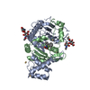

 SPODOPTERA FRUGIPERDA (fall armyworm) / References: UniProt: P10619, carboxypeptidase C


 Mass: 383.373 Da / Num. of mol.: 1 / Source method: obtained synthetically / Formula: C20H18FN3O4
 Mass: 112.411 Da / Num. of mol.: 6 / Source method: obtained synthetically / Formula: Cd
 Sample preparation
 / Beamline: ID29 / Wavelength: 0.9762
  Processing