 Movie
Movie Controller


+ Open data

- Basic information
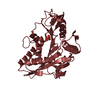
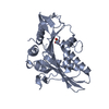
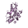
| Entry | Database: PDB / ID: 3x2t | ||||||
|---|---|---|---|---|---|---|---|
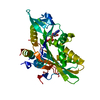
| Title | Crystal Structure of the KIF5C Motor Domain With ADP | ||||||
 Components Components | Kinesin heavy chain isoform 5C | ||||||
 Keywords Keywords | MOTOR PROTEIN / kinesin / motor domain / ATPase / nucleotide binding / microtubule / transport protein | ||||||
| Function / homology |  Function and homology information Function and homology informationdistal axon / anterograde dendritic transport of messenger ribonucleoprotein complex / anterograde dendritic transport of neurotransmitter receptor complex / anterograde axonal protein transport / apolipoprotein receptor binding / ciliary rootlet / intracellular mRNA localization / plus-end-directed microtubule motor activity / motor neuron axon guidance / microtubule motor activity ...distal axon / anterograde dendritic transport of messenger ribonucleoprotein complex / anterograde dendritic transport of neurotransmitter receptor complex / anterograde axonal protein transport / apolipoprotein receptor binding / ciliary rootlet / intracellular mRNA localization / plus-end-directed microtubule motor activity / motor neuron axon guidance / microtubule motor activity / kinesin complex / synaptic vesicle transport / mRNA transport / postsynaptic cytosol / axonal growth cone / axon cytoplasm / dendrite cytoplasm / axon guidance / Hydrolases; Acting on acid anhydrides; Acting on acid anhydrides to facilitate cellular and subcellular movement / GABA-ergic synapse / microtubule binding / microtubule / neuron projection / neuronal cell body / ATP hydrolysis activity / ATP binding / cytoplasm Similarity search - Function | ||||||
| Biological species |  | ||||||
| Method |  X-RAY DIFFRACTION / SYNCHROTRON / MOLECULAR REPLACEMENT / Resolution: 2.7 Å X-RAY DIFFRACTION / SYNCHROTRON / MOLECULAR REPLACEMENT / Resolution: 2.7 Å | ||||||
 Authors Authors | Inoue, S. / Nitta, R. / Hirokawa, N. | ||||||
 Citation Citation | Journal: EMBO J / Year: 2015 Title: X-ray and Cryo-EM structures reveal mutual conformational changes of Kinesin and GTP-state microtubules upon binding. Authors: Manatsu Morikawa / Hiroaki Yajima / Ryo Nitta / Shigeyuki Inoue / Toshihiko Ogura / Chikara Sato / Nobutaka Hirokawa /   Abstract: The molecular motor kinesin moves along microtubules using energy from ATP hydrolysis in an initial step coupled with ADP release. In neurons, kinesin-1/KIF5C preferentially binds to the GTP-state ...The molecular motor kinesin moves along microtubules using energy from ATP hydrolysis in an initial step coupled with ADP release. In neurons, kinesin-1/KIF5C preferentially binds to the GTP-state microtubules over GDP-state microtubules to selectively enter an axon among many processes; however, because the atomic structure of nucleotide-free KIF5C is unavailable, its molecular mechanism remains unresolved. Here, the crystal structure of nucleotide-free KIF5C and the cryo-electron microscopic structure of nucleotide-free KIF5C complexed with the GTP-state microtubule are presented. The structures illustrate mutual conformational changes induced by interaction between the GTP-state microtubule and KIF5C. KIF5C acquires the 'rigor conformation', where mobile switches I and II are stabilized through L11 and the initial portion of the neck-linker, facilitating effective ADP release and the weak-to-strong transition of KIF5C microtubule affinity. Conformational changes to tubulin strengthen the longitudinal contacts of the GTP-state microtubule in a similar manner to GDP-taxol microtubules. These results and functional analyses provide the molecular mechanism of the preferential binding of KIF5C to GTP-state microtubules. | ||||||
| History |
|
- Structure visualization
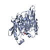
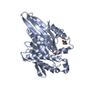
| Structure viewer | Molecule: MolmilJmol/JSmol |
|---|
- Downloads & links
-Download
| PDBx/mmCIF format | 3x2t.cif.gz | 139.4 KB | Display | PDBx/mmCIF format |
|---|---|---|---|---|
| PDB format | pdb3x2t.ent.gz | 108.9 KB | Display | PDB format |
| PDBx/mmJSON format | 3x2t.json.gz | Tree view | PDBx/mmJSON format | |
| Others |  Other downloads Other downloads |
-Validation report
| Arichive directory | https://data.pdbj.org/pub/pdb/validation_reports/x2/3x2tftp://data.pdbj.org/pub/pdb/validation_reports/x2/3x2t | HTTPS FTP |
|---|
-Related structure data
| Related structure data | 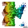 5916C  3j6hC  3wrdC  2kinS C: citing same article ( S: Starting model for refinement |
|---|---|
| Similar structure data |


-Links
 PDBj
PDBj


- Assembly
| Deposited unit | 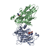
| ||||||||
|---|---|---|---|---|---|---|---|---|---|
| 1 | 
| ||||||||
| 2 | 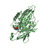
| ||||||||
| Unit cell |
|
-Components
| #1: Protein | Mass: 38255.551 Da / Num. of mol.: 2 / Fragment: Motor domain, RESIDUES 1-334 Source method: isolated from a genetically manipulated source Source: (gene. exp.)  #2: Chemical |   Mass: 427.201 Da / Num. of mol.: 2 / Source method: obtained synthetically / Formula: C10H15N5O10P2 / Comment: ADP, energy-carrying molecule*YM Mass: 427.201 Da / Num. of mol.: 2 / Source method: obtained synthetically / Formula: C10H15N5O10P2 / Comment: ADP, energy-carrying molecule*YM#3: Water | ChemComp-HOH / |  Mass: 18.015 Da / Num. of mol.: 5 / Source method: isolated from a natural source / Formula: H2O Mass: 18.015 Da / Num. of mol.: 5 / Source method: isolated from a natural source / Formula: H2O |
|---|
-Experimental details
-Experiment
| Experiment | Method: X-RAY DIFFRACTION / Number of used crystals: 1 |
|---|
- Sample preparation
| Crystal | Density Matthews: 2.98 Å3/Da / Density % sol: 58.77 % / Mosaicity: 0.597 ° / Mosaicity esd: 0.007 ° |
|---|---|
| Crystal grow | Temperature: 293 K / Method: vapor diffusion, hanging drop / pH: 5.5 Details: Ammonium sulfate, Sodium citrate, Glycerol, pH 5.5, VAPOR DIFFUSION, HANGING DROP, temperature 293K |
-Data collection
| Diffraction | Mean temperature: 95 K | ||||||||||||||||||||||||||||||||||||||||||||||||||||||||||||||||||
|---|---|---|---|---|---|---|---|---|---|---|---|---|---|---|---|---|---|---|---|---|---|---|---|---|---|---|---|---|---|---|---|---|---|---|---|---|---|---|---|---|---|---|---|---|---|---|---|---|---|---|---|---|---|---|---|---|---|---|---|---|---|---|---|---|---|---|---|
| Diffraction source | Source: SYNCHROTRON / Site: Photon Factory / Beamline: AR-NW12A / Wavelength: 1 Å | ||||||||||||||||||||||||||||||||||||||||||||||||||||||||||||||||||
| Detector | Type: ADSC QUANTUM 210 / Detector: CCD / Date: Dec 21, 2008 | ||||||||||||||||||||||||||||||||||||||||||||||||||||||||||||||||||
| Radiation | Monochromator: Si(111) / Protocol: SINGLE WAVELENGTH / Monochromatic (M) / Laue (L): M / Scattering type: x-ray | ||||||||||||||||||||||||||||||||||||||||||||||||||||||||||||||||||
| Radiation wavelength | Wavelength: 1 Å / Relative weight: 1 | ||||||||||||||||||||||||||||||||||||||||||||||||||||||||||||||||||
| Reflection | Resolution: 2.7→50 Å / Num. all: 25884 / Num. obs: 25753 / % possible obs: 99.5 % / Redundancy: 6.8 % / Rmerge(I) obs: 0.049 / Χ2: 0.683 / Net I/σ(I): 14.6 | ||||||||||||||||||||||||||||||||||||||||||||||||||||||||||||||||||
| Reflection shell | Diffraction-ID: 1 / Rejects: _
|
- Processing
| Software |
| |||||||||||||||||||||||||||||||||||||||||||||||||||||||||||||||||||||||||||||||||||||||||||||||||||||||||||||||||||||||||||||||||||||||||||||||||||
|---|---|---|---|---|---|---|---|---|---|---|---|---|---|---|---|---|---|---|---|---|---|---|---|---|---|---|---|---|---|---|---|---|---|---|---|---|---|---|---|---|---|---|---|---|---|---|---|---|---|---|---|---|---|---|---|---|---|---|---|---|---|---|---|---|---|---|---|---|---|---|---|---|---|---|---|---|---|---|---|---|---|---|---|---|---|---|---|---|---|---|---|---|---|---|---|---|---|---|---|---|---|---|---|---|---|---|---|---|---|---|---|---|---|---|---|---|---|---|---|---|---|---|---|---|---|---|---|---|---|---|---|---|---|---|---|---|---|---|---|---|---|---|---|---|---|---|---|---|
| Refinement | Method to determine structure: MOLECULAR REPLACEMENT Starting model: PDB ENTRY 2KIN Resolution: 2.7→20 Å / Cor.coef. Fo:Fc: 0.942 / Cor.coef. Fo:Fc free: 0.881 / WRfactor Rfree: 0.303 / WRfactor Rwork: 0.228 / SU B: 15.697 / SU ML: 0.323 / Cross valid method: THROUGHOUT / σ(F): 0 / ESU R: 0.685 / ESU R Free: 0.374 / Stereochemistry target values: MAXIMUM LIKELIHOOD Details: HYDROGENS HAVE BEEN ADDED IN THE RIDING POSITIONS U VALUES: REFINED INDIVIDUALLY
| |||||||||||||||||||||||||||||||||||||||||||||||||||||||||||||||||||||||||||||||||||||||||||||||||||||||||||||||||||||||||||||||||||||||||||||||||||
| Solvent computation | Ion probe radii: 0.8 Å / Shrinkage radii: 0.8 Å / VDW probe radii: 1.2 Å / Solvent model: MASK | |||||||||||||||||||||||||||||||||||||||||||||||||||||||||||||||||||||||||||||||||||||||||||||||||||||||||||||||||||||||||||||||||||||||||||||||||||
| Displacement parameters | Biso max: 200.98 Å2 / Biso mean: 84.422 Å2 / Biso min: 38.05 Å2
| |||||||||||||||||||||||||||||||||||||||||||||||||||||||||||||||||||||||||||||||||||||||||||||||||||||||||||||||||||||||||||||||||||||||||||||||||||
| Refinement step | Cycle: LAST / Resolution: 2.7→20 Å
| |||||||||||||||||||||||||||||||||||||||||||||||||||||||||||||||||||||||||||||||||||||||||||||||||||||||||||||||||||||||||||||||||||||||||||||||||||
| Refine LS restraints |
| |||||||||||||||||||||||||||||||||||||||||||||||||||||||||||||||||||||||||||||||||||||||||||||||||||||||||||||||||||||||||||||||||||||||||||||||||||
| LS refinement shell | Refine-ID: X-RAY DIFFRACTION / Total num. of bins used: 20
|
