[English] 日本語
 Yorodumi
- EMDB-5916: Cryo-Electron Microscopy of Nucleotide-free Kinesin motor domain ... -
+ Open data
- Basic information
| Entry | Database: EMDB / ID: EMD-5916 | |||||||||
|---|---|---|---|---|---|---|---|---|---|---|
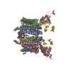
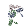
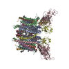
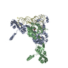
| Title | Cryo-Electron Microscopy of Nucleotide-free Kinesin motor domain complexed with GMPCPP-microtubule | |||||||||
 Map data Map data | 3D structure of Nucleotide-free Kinesin motor domain complexed with GMPCPP-microtubule | |||||||||
 Sample Sample |
| |||||||||
 Keywords Keywords | KIF5C / nucleotide-free kinesin / Rigor-conformation / GMPCPP-MT / Axonal transport / transport protein | |||||||||
| Function / homology |  Function and homology information Function and homology informationdistal axon / anterograde dendritic transport of messenger ribonucleoprotein complex / anterograde axonal protein transport / apolipoprotein receptor binding / intracellular mRNA localization / ciliary rootlet / motor neuron axon guidance / kinesin complex / microtubule motor activity / motile cilium ...distal axon / anterograde dendritic transport of messenger ribonucleoprotein complex / anterograde axonal protein transport / apolipoprotein receptor binding / intracellular mRNA localization / ciliary rootlet / motor neuron axon guidance / kinesin complex / microtubule motor activity / motile cilium / mRNA transport / postsynaptic cytosol / axonal growth cone / axon cytoplasm / dendrite cytoplasm / structural constituent of cytoskeleton / GABA-ergic synapse / Hydrolases; Acting on acid anhydrides; Acting on acid anhydrides to facilitate cellular and subcellular movement / microtubule cytoskeleton organization / neuron migration / mitotic cell cycle / microtubule binding / Hydrolases; Acting on acid anhydrides; Acting on GTP to facilitate cellular and subcellular movement / microtubule / neuron projection / hydrolase activity / neuronal cell body / GTPase activity / GTP binding / ATP hydrolysis activity / ATP binding / metal ion binding / cytoplasm Similarity search - Function | |||||||||
| Biological species |   | |||||||||
| Method | single particle reconstruction / cryo EM / negative staining / Resolution: 8.1 Å | |||||||||
 Authors Authors | Morikawa M / Yajima H / Nitta R / Inoue S / Ogura T / Sato C / Hirokawa N | |||||||||
 Citation Citation |  Journal: EMBO J / Year: 2015 Journal: EMBO J / Year: 2015Title: X-ray and Cryo-EM structures reveal mutual conformational changes of Kinesin and GTP-state microtubules upon binding. Authors: Manatsu Morikawa / Hiroaki Yajima / Ryo Nitta / Shigeyuki Inoue / Toshihiko Ogura / Chikara Sato / Nobutaka Hirokawa /   Abstract: The molecular motor kinesin moves along microtubules using energy from ATP hydrolysis in an initial step coupled with ADP release. In neurons, kinesin-1/KIF5C preferentially binds to the GTP-state ...The molecular motor kinesin moves along microtubules using energy from ATP hydrolysis in an initial step coupled with ADP release. In neurons, kinesin-1/KIF5C preferentially binds to the GTP-state microtubules over GDP-state microtubules to selectively enter an axon among many processes; however, because the atomic structure of nucleotide-free KIF5C is unavailable, its molecular mechanism remains unresolved. Here, the crystal structure of nucleotide-free KIF5C and the cryo-electron microscopic structure of nucleotide-free KIF5C complexed with the GTP-state microtubule are presented. The structures illustrate mutual conformational changes induced by interaction between the GTP-state microtubule and KIF5C. KIF5C acquires the 'rigor conformation', where mobile switches I and II are stabilized through L11 and the initial portion of the neck-linker, facilitating effective ADP release and the weak-to-strong transition of KIF5C microtubule affinity. Conformational changes to tubulin strengthen the longitudinal contacts of the GTP-state microtubule in a similar manner to GDP-taxol microtubules. These results and functional analyses provide the molecular mechanism of the preferential binding of KIF5C to GTP-state microtubules. | |||||||||
| History |
|
- Structure visualization
| Movie |
 Movie viewer Movie viewer |
|---|---|
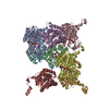
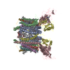
| Structure viewer | EM map:  SurfView SurfView Molmil Molmil Jmol/JSmol Jmol/JSmol |
| Supplemental images |
- Downloads & links
-EMDB archive
| Map data |  emd_5916.map.gz emd_5916.map.gz | 179.5 KB |  EMDB map data format EMDB map data format | |
|---|---|---|---|---|
| Header (meta data) |  emd-5916-v30.xml emd-5916-v30.xml emd-5916.xml emd-5916.xml | 13.2 KB 13.2 KB | Display Display |  EMDB header EMDB header |
| Images |  emd_5916.tif emd_5916.tif | 316.3 KB | ||
| Archive directory |  http://ftp.pdbj.org/pub/emdb/structures/EMD-5916 http://ftp.pdbj.org/pub/emdb/structures/EMD-5916 ftp://ftp.pdbj.org/pub/emdb/structures/EMD-5916 ftp://ftp.pdbj.org/pub/emdb/structures/EMD-5916 | HTTPS FTP |
-Validation report
| Summary document |  emd_5916_validation.pdf.gz emd_5916_validation.pdf.gz | 308.6 KB | Display |  EMDB validaton report EMDB validaton report |
|---|---|---|---|---|
| Full document |  emd_5916_full_validation.pdf.gz emd_5916_full_validation.pdf.gz | 308.1 KB | Display | |
| Data in XML |  emd_5916_validation.xml.gz emd_5916_validation.xml.gz | 4.3 KB | Display | |
| Arichive directory |  https://ftp.pdbj.org/pub/emdb/validation_reports/EMD-5916 https://ftp.pdbj.org/pub/emdb/validation_reports/EMD-5916 ftp://ftp.pdbj.org/pub/emdb/validation_reports/EMD-5916 ftp://ftp.pdbj.org/pub/emdb/validation_reports/EMD-5916 | HTTPS FTP |
-Related structure data
| Related structure data |  3j6hMC  3wrdC  3x2tC M: atomic model generated by this map C: citing same article ( |
|---|---|
| Similar structure data |
- Links
Links
| EMDB pages |  EMDB (EBI/PDBe) / EMDB (EBI/PDBe) /  EMDataResource EMDataResource |
|---|---|
| Related items in Molecule of the Month |
- Map
Map
| File |  Download / File: emd_5916.map.gz / Format: CCP4 / Size: 199.2 KB / Type: IMAGE STORED AS FLOATING POINT NUMBER (4 BYTES) Download / File: emd_5916.map.gz / Format: CCP4 / Size: 199.2 KB / Type: IMAGE STORED AS FLOATING POINT NUMBER (4 BYTES) | ||||||||||||||||||||||||||||||||||||||||||||||||||||||||||||
|---|---|---|---|---|---|---|---|---|---|---|---|---|---|---|---|---|---|---|---|---|---|---|---|---|---|---|---|---|---|---|---|---|---|---|---|---|---|---|---|---|---|---|---|---|---|---|---|---|---|---|---|---|---|---|---|---|---|---|---|---|---|
| Annotation | 3D structure of Nucleotide-free Kinesin motor domain complexed with GMPCPP-microtubule | ||||||||||||||||||||||||||||||||||||||||||||||||||||||||||||
| Projections & slices | Image control
Images are generated by Spider. generated in cubic-lattice coordinate | ||||||||||||||||||||||||||||||||||||||||||||||||||||||||||||
| Voxel size | X=Y=Z: 2.5 Å | ||||||||||||||||||||||||||||||||||||||||||||||||||||||||||||
| Density |
| ||||||||||||||||||||||||||||||||||||||||||||||||||||||||||||
| Symmetry | Space group: 1 | ||||||||||||||||||||||||||||||||||||||||||||||||||||||||||||
| Details | EMDB XML:
CCP4 map header:
| ||||||||||||||||||||||||||||||||||||||||||||||||||||||||||||
-Supplemental data
- Sample components
-Entire : Cryo-Electron Microscopy of Nucleotide-free Kinesin motor domain ...
| Entire | Name: Cryo-Electron Microscopy of Nucleotide-free Kinesin motor domain complexed with GMPCPP-microtubule |
|---|---|
| Components |
|
-Supramolecule #1000: Cryo-Electron Microscopy of Nucleotide-free Kinesin motor domain ...
| Supramolecule | Name: Cryo-Electron Microscopy of Nucleotide-free Kinesin motor domain complexed with GMPCPP-microtubule type: sample / ID: 1000 Oligomeric state: One kineisn motor domain binds to one tubulin dimers Number unique components: 3 |
|---|
-Macromolecule #1: TUBULIN ALPHA CHAIN
| Macromolecule | Name: TUBULIN ALPHA CHAIN / type: protein_or_peptide / ID: 1 / Name.synonym: alpha tubulin / Number of copies: 1 / Recombinant expression: No / Database: NCBI |
|---|---|
| Source (natural) | Organism:  |
| Sequence | UniProtKB: Tubulin alpha-1A chain |
-Macromolecule #2: TUBULIN BETA CHAIN
| Macromolecule | Name: TUBULIN BETA CHAIN / type: protein_or_peptide / ID: 2 / Name.synonym: beta tubulin / Number of copies: 1 / Recombinant expression: No / Database: NCBI |
|---|---|
| Source (natural) | Organism:  |
| Sequence | UniProtKB: Tubulin beta chain |
-Macromolecule #3: Kinesin heavy chain isoform 5C motor domain
| Macromolecule | Name: Kinesin heavy chain isoform 5C motor domain / type: protein_or_peptide / ID: 3 / Name.synonym: KIF5C motor domain / Number of copies: 1 / Recombinant expression: Yes |
|---|---|
| Source (natural) | Organism:  |
| Recombinant expression | Organism:  |
| Sequence | UniProtKB: Kinesin heavy chain isoform 5C |
-Experimental details
-Structure determination
| Method | negative staining, cryo EM |
|---|---|
 Processing Processing | single particle reconstruction |
| Aggregation state | particle |
- Sample preparation
| Buffer | pH: 6.8 / Details: 100mM PIPES, 1mM EGTA, 1mM MgCl2 |
|---|---|
| Staining | Type: NEGATIVE / Details: No staining |
| Grid | Details: 300 mesh copper grid with thin carbon support, glow discharged in amylamine atmosphere |
| Vitrification | Cryogen name: ETHANE / Chamber temperature: 100 K / Instrument: LEICA KF80 / Method: On grid blotting (Microtubule, KIF5C) |
- Electron microscopy
| Microscope | JEOL 2010F |
|---|---|
| Date | Aug 18, 2012 |
| Image recording | Category: FILM / Film or detector model: KODAK SO-163 FILM / Digitization - Scanner: OTHER / Average electron dose: 7 e/Å2 |
| Tilt angle min | 0 |
| Tilt angle max | 0 |
| Electron beam | Acceleration voltage: 200 kV / Electron source:  FIELD EMISSION GUN FIELD EMISSION GUN |
| Electron optics | Calibrated magnification: 40000 / Illumination mode: FLOOD BEAM / Imaging mode: BRIGHT FIELD / Nominal defocus max: 2.6 µm / Nominal defocus min: 1.2 µm / Nominal magnification: 40000 |
| Sample stage | Specimen holder: Liquid nitrogen cooled / Specimen holder model: GATAN LIQUID NITROGEN |
- Image processing
| Details | The particles were selected along individual microtubules using an automatic processing program. |
|---|---|
| CTF correction | Details: Each filament |
| Final reconstruction | Algorithm: OTHER / Resolution.type: BY AUTHOR / Resolution: 8.1 Å / Resolution method: OTHER / Software - Name: MATLAB, ImagicV / Number images used: 302000 |
| Final two d classification | Number classes: 18 |
-Atomic model buiding 1
| Initial model | PDB ID: Chain - #0 - Chain ID: A / Chain - #1 - Chain ID: B |
|---|---|
| Software | Name: Chimera, Modeller |
| Details | Initial local fitting was done using Chimera, and for some loops Modeller was used. |
| Refinement | Space: REAL / Protocol: RIGID BODY FIT Target criteria: Cross-correlation, Average map value, Atoms inside the contour |
| Output model |  PDB-3j6h: |
-Atomic model buiding 2
| Initial model | PDB ID: Chain - Chain ID: K |
|---|---|
| Software | Name: Chimera, Modeller |
| Details | Initial local fitting was done using Chimera, and for some loops Modeller was used. |
| Refinement | Space: REAL / Protocol: RIGID BODY FIT Target criteria: Cross-correlation, Average map value, Atoms inside the contour |
| Output model |  PDB-3j6h: |
 Movie
Movie Controller






 Z (Sec.)
 Y (Row.)
 X (Col.)


