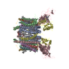
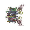
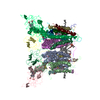
Method to determine structure: MOLECULAR REPLACEMENT / Resolution: 3.41→41.27 Å / Cor.coef. Fo:Fc: 0.922 / Cor.coef. Fo:Fc free: 0.86 / SU B: 15.94 / SU ML: 0.243 / TLS residual ADP flag: LIKELY RESIDUAL / Cross valid method: THROUGHOUT / ESU R: 1.115 / ESU R Free: 0.426 / Stereochemistry target values: MAXIMUM LIKELIHOOD Details: HYDROGENS HAVE BEEN ADDED IN THE RIDING POSITIONS. Many of the basic and novel features of the structure of the cyanobacterial b6f complex reported now in entries 2E74 (native), 2E75 (with ...Details: HYDROGENS HAVE BEEN ADDED IN THE RIDING POSITIONS. Many of the basic and novel features of the structure of the cyanobacterial b6f complex reported now in entries 2E74 (native), 2E75 (with quinone analogue inhibitor NQNO), and 2E76 (with quinone analogue inhibitor TDS) were seen in the original 3.0 A structure that was refined in space group P61 (Science, 302:1009-, 2003; pdb entry, 1VF5). This structure was thought to be a co-complex with tridecyl-stigmatellin (TDS). This inference was based on: (i) the highest resolution of 3 A was obtained in the TDS co-crystals, the native structure having a poorer resolution; (ii) electron density outside the portal on the p-side of the quinone exchange cavity resembled the TDS ring. Because of the poorer resolution of the native complex at that time, it was not possible to check for the presence of this density in the native structure. Entry 2E74 reports a 3.0 A native structure obtained in the presence of Cd2+, which shows the density previously attributed to the TDS ring. The correct p-side position of TDS, reported in 2E76, and in agreement with its location in the C. reinhardtii b6f structure (entry 1Q90) was obtained when the DOPC lipid that was added to accelerate crystallization (PNAS,100: 5160-5163) was added after TDS. 2E76 also shows a unique second binding site for TDS on the n-side of the complex, close to the position of an axial ligand of heme cn. Entry 2E75 shows that the inhibitor NQNO occupies a similar n-side binding site. This site that is common to the binding of the two quinone analogue inhibitors implies that it is also the n-side binding site of plastoquinone. 2E74,2E75, and 2E76 were refined in space group P6122.
Rfactor
Num. reflection
% reflection
Selection details
Rfree
0.25563
1852
5 %
RANDOM
Rwork
0.18473
-
-
-
obs
0.18822
35129
99.68 %
-
Solvent computation
Ion probe radii: 0.8 Å / Shrinkage radii: 0.8 Å / VDW probe radii: 1.4 Å / Solvent model: MASK
Displacement parameters
Biso mean: 62.768 Å2
Baniso -1
Baniso -2
Baniso -3
1-
1.02 Å2
0.51 Å2
0 Å2
2-
-
1.02 Å2
0 Å2
3-
-
-
-1.53 Å2
Refinement step
Cycle: LAST / Resolution: 3.41→41.27 Å
Protein
Nucleic acid
Ligand
Solvent
Total
Num. atoms
7467
0
640
5
8112
Refine LS restraints
Refine-ID
Type
Dev ideal
Dev ideal target
Number
X-RAY DIFFRACTION
r_bond_refined_d
0.037
0.022
8340
X-RAY DIFFRACTION
r_bond_other_d
X-RAY DIFFRACTION
r_angle_refined_deg
4.042
2.089
11387
X-RAY DIFFRACTION
r_angle_other_deg
X-RAY DIFFRACTION
r_dihedral_angle_1_deg
12.51
5
952
X-RAY DIFFRACTION
r_dihedral_angle_2_deg
40.162
24.164
293
X-RAY DIFFRACTION
r_dihedral_angle_3_deg
27.18
15
1246
X-RAY DIFFRACTION
r_dihedral_angle_4_deg
25.672
15
26
X-RAY DIFFRACTION
r_chiral_restr
0.242
0.2
1250
X-RAY DIFFRACTION
r_gen_planes_refined
0.012
0.02
6135
X-RAY DIFFRACTION
r_gen_planes_other
X-RAY DIFFRACTION
r_nbd_refined
0.397
0.2
5047
X-RAY DIFFRACTION
r_nbd_other
X-RAY DIFFRACTION
r_nbtor_refined
0.386
0.2
5482
X-RAY DIFFRACTION
r_nbtor_other
X-RAY DIFFRACTION
r_xyhbond_nbd_refined
0.249
0.2
398
X-RAY DIFFRACTION
r_xyhbond_nbd_other
X-RAY DIFFRACTION
r_metal_ion_refined
0.235
0.2
7
X-RAY DIFFRACTION
r_metal_ion_other
X-RAY DIFFRACTION
r_symmetry_vdw_refined
0.33
0.2
93
X-RAY DIFFRACTION
r_symmetry_vdw_other
X-RAY DIFFRACTION
r_symmetry_hbond_refined
0.37
0.2
3
X-RAY DIFFRACTION
r_symmetry_hbond_other
X-RAY DIFFRACTION
r_symmetry_metal_ion_refined
X-RAY DIFFRACTION
r_symmetry_metal_ion_other
X-RAY DIFFRACTION
r_mcbond_it
6.938
1.5
4944
X-RAY DIFFRACTION
r_mcbond_other
X-RAY DIFFRACTION
r_mcangle_it
10.05
2
7739
X-RAY DIFFRACTION
r_scbond_it
16.754
3
4250
X-RAY DIFFRACTION
r_scangle_it
21.711
4.5
3641
X-RAY DIFFRACTION
r_rigid_bond_restr
X-RAY DIFFRACTION
r_sphericity_free
X-RAY DIFFRACTION
r_sphericity_bonded
LS refinement shell
Resolution: 3.408→3.496 Å / Total num. of bins used: 20
Rfactor
Num. reflection
% reflection
Rfree
0.296
120
-
Rwork
0.21
2476
-
obs
-
-
96.97 %
Refinement TLS params.
Method: refined / Refine-ID: X-RAY DIFFRACTION
ID
L11 (°2)
L12 (°2)
L13 (°2)
L22 (°2)
L23 (°2)
L33 (°2)
S11 (Å °)
S12 (Å °)
S13 (Å °)
S21 (Å °)
S22 (Å °)
S23 (Å °)
S31 (Å °)
S32 (Å °)
S33 (Å °)
T11 (Å2)
T12 (Å2)
T13 (Å2)
T22 (Å2)
T23 (Å2)
T33 (Å2)
Origin x (Å)
Origin y (Å)
Origin z (Å)
1
5.0059
-1.8657
2.0697
8.1587
-0.1308
2.702
-0.3365
-0.521
-0.2702
1.6895
0.8479
1.0223
-1.1058
-0.533
-0.5115
0.3519
0.2707
0.4967
-0.0583
0.1973
0.0879
-74.5308
75.2426
55.1095
2
3.1496
1.9643
2.1113
6.401
-1.437
3.7304
0.5099
-0.0633
-0.3485
-0.0125
-0.0218
2.1133
-0.178
-0.8957
-0.4882
-0.2874
0.182
0.2929
-0.0402
0.0767
0.6755
-84.3264
69.8315
44.6672
3
2.6815
2.3661
-3.0986
5.3203
-3.9032
4.0034
0.2059
-0.3697
0.7364
0.2309
-0.2739
0.1917
-0.3569
0.1443
0.068
-0.0101
-0.0271
-0.0082
-0.1101
0.1264
0.1327
-44.6738
93.8622
17.7702
4
2.0917
1.6383
-0.8558
3.5575
-3.3115
3.4172
0.1853
0.0877
-1.0223
-1.0791
-0.2839
-0.3928
0.898
0.1905
0.0986
0.3858
0.3605
-0.3941
0.2043
-0.7213
0.3237
-64.166
19.4696
-19.5406
5
2.128
0.0551
0.4816
2.4156
0.8975
1.1772
0.2218
0.7416
-0.0263
-0.0363
-0.0031
0.1242
0.0739
-0.0487
-0.2188
-0.1507
0.0969
-0.0811
0.3389
-0.1096
-0.1004
-69.1522
55.6639
-7.2531
6
3.0178
2.8308
-0.5672
2.7826
-0.3735
1.0459
0.1035
0.3295
0.3968
0.0182
-0.0809
0.0471
-0.4443
0.0214
-0.0226
-0.0121
-0.0309
0.0361
-0.02
0.21
0.1743
-39.3888
93.9693
7.9105
Refinement TLS group
ID
Refine-ID
Refine TLS-ID
Auth asym-ID
Auth seq-ID
1
X-RAY DIFFRACTION
1
D
100 - 148
2
X-RAY DIFFRACTION
2
D
54 - 99
3
X-RAY DIFFRACTION
2
D
149 - 179
4
X-RAY DIFFRACTION
3
D
9 - 46
5
X-RAY DIFFRACTION
4
C
171 - 233
6
X-RAY DIFFRACTION
5
C
1 - 169
7
X-RAY DIFFRACTION
5
C
236 - 251
8
X-RAY DIFFRACTION
6
C
253 - 288
+
About Yorodumi
-
News
-
Feb 9, 2022. New format data for meta-information of EMDB entries
New format data for meta-information of EMDB entries
Version 3 of the EMDB header file is now the official format.
The previous official version 1.9 will be removed from the archive.
In the structure databanks used in Yorodumi, some data are registered as the other names, "COVID-19 virus" and "2019-nCoV". Here are the details of the virus and the list of structure data.
Jan 31, 2019. EMDB accession codes are about to change! (news from PDBe EMDB page)
EMDB accession codes are about to change! (news from PDBe EMDB page)
The allocation of 4 digits for EMDB accession codes will soon come to an end. Whilst these codes will remain in use, new EMDB accession codes will include an additional digit and will expand incrementally as the available range of codes is exhausted. The current 4-digit format prefixed with “EMD-” (i.e. EMD-XXXX) will advance to a 5-digit format (i.e. EMD-XXXXX), and so on. It is currently estimated that the 4-digit codes will be depleted around Spring 2019, at which point the 5-digit format will come into force.
The EM Navigator/Yorodumi systems omit the EMD- prefix.
Related info.:Q: What is EMD? / ID/Accession-code notation in Yorodumi/EM Navigator
Yorodumi is a browser for structure data from EMDB, PDB, SASBDB, etc.
This page is also the successor to EM Navigator detail page, and also detail information page/front-end page for Omokage search.
The word "yorodu" (or yorozu) is an old Japanese word meaning "ten thousand". "mi" (miru) is to see.
Related info.:EMDB / PDB / SASBDB / Comparison of 3 databanks / Yorodumi Search / Aug 31, 2016. New EM Navigator & Yorodumi / Yorodumi Papers / Jmol/JSmol / Function and homology information / Changes in new EM Navigator and Yorodumi
 Movie
Movie Controller

 Yorodumi
 Open data
 Basic information
 Components
 Keywords
 Function and homology information
 Mastigocladus laminosus (bacteria)
 X-RAY DIFFRACTION /
  Authors
 Citation
 Structure visualization
 Downloads & links
 Other downloads
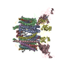


 PDBj
PDBj


 Assembly


 Mass: 112.411 Da / Num. of mol.: 1 / Source method: obtained synthetically / Formula: Cd
 Mass: 616.487 Da / Num. of mol.: 2 / Source method: obtained synthetically / Formula: C34H32FeN4O4
 Mass: 618.503 Da / Num. of mol.: 2 / Source method: obtained synthetically / Formula: C34H34FeN4O4
 Mass: 801.148 Da / Num. of mol.: 2 / Source method: obtained synthetically / Formula: C45H87NO8P / Comment: phospholipid*YM
 Mass: 496.589 Da / Num. of mol.: 4 / Source method: obtained synthetically / Formula: C23H44O11 / Comment: detergent*YM
 Mass: 893.489 Da / Num. of mol.: 1 / Source method: obtained synthetically / Formula: C55H72MgN4O5
 Mass: 418.566 Da / Num. of mol.: 2 / Source method: obtained synthetically / Formula: C25H38O5
 Mass: 175.820 Da / Num. of mol.: 1 / Source method: obtained synthetically / Formula: Fe2S2
 Mass: 795.116 Da / Num. of mol.: 1 / Source method: obtained synthetically / Formula: C41H78O12S
 Mass: 536.873 Da / Num. of mol.: 1 / Source method: obtained synthetically / Formula: C40H56
 Sample preparation
 / Beamline: 19-ID / Wavelength: 0.97856 Å
 Processing