 Movie
Movie Controller

[English] 日本語
 Yorodumi
- PDB-4mea: Crystal structure of the Cif epoxide hydrolase from Acinetobacter... -
+ Open data

- Basic information
| Entry | Database: PDB / ID: 4mea | ||||||
|---|---|---|---|---|---|---|---|
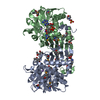
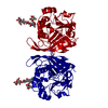
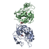
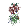
| Title | Crystal structure of the Cif epoxide hydrolase from Acinetobacter nosocomialis | ||||||
 Components Components | Predicted protein | ||||||
 Keywords Keywords | HYDROLASE / alpha/beta hydrolase fold / epoxide hydrolase / secreted | ||||||
| Function / homology | Alpha/Beta hydrolase fold, catalytic domain / Rossmann fold / 3-Layer(aba) Sandwich / Alpha Beta / :  Function and homology information Function and homology information | ||||||
| Biological species |  Acinetobacter sp. RUH2624 (bacteria) Acinetobacter sp. RUH2624 (bacteria) | ||||||
| Method |  X-RAY DIFFRACTION / SYNCHROTRON / MOLECULAR REPLACEMENT / Resolution: 1.95 Å X-RAY DIFFRACTION / SYNCHROTRON / MOLECULAR REPLACEMENT / Resolution: 1.95 Å | ||||||
 Authors Authors | Bahl, C.D. / Madden, D.R. | ||||||
 Citation Citation | Journal: J.Biol.Chem. / Year: 2014 Title: Signature motifs identify an acinetobacter cif virulence factor with epoxide hydrolase activity. Authors: Bahl, C.D. / Hvorecny, K.L. / Bridges, A.A. / Ballok, A.E. / Bomberger, J.M. / Cady, K.C. / O'Toole, G.A. / Madden, D.R. | ||||||
| History |
|
- Structure visualization
| Structure viewer | Molecule: MolmilJmol/JSmol |
|---|
- Downloads & links
-Download
| PDBx/mmCIF format | 4mea.cif.gz | 252.9 KB | Display | PDBx/mmCIF format |
|---|---|---|---|---|
| PDB format | pdb4mea.ent.gz | 202.1 KB | Display | PDB format |
| PDBx/mmJSON format | 4mea.json.gz | Tree view | PDBx/mmJSON format | |
| Others |  Other downloads Other downloads |
-Validation report
| Arichive directory | https://data.pdbj.org/pub/pdb/validation_reports/me/4meaftp://data.pdbj.org/pub/pdb/validation_reports/me/4mea | HTTPS FTP |
|---|
-Related structure data
| Related structure data |  4mebC  3kd2S S: Starting model for refinement C: citing same article ( |
|---|---|
| Similar structure data |


-Links
 PDBj
PDBj- Assembly
| Deposited unit | 
| ||||||||
|---|---|---|---|---|---|---|---|---|---|
| 1 |
| ||||||||
| Unit cell |
|
-Components
| #1: Protein | Mass: 36857.859 Da / Num. of mol.: 2 / Fragment: UNP residues 25-349 Source method: isolated from a genetically manipulated source Source: (gene. exp.) Acinetobacter sp. RUH2624 (bacteria) / Gene: acif, HMPREF0014_00517 / Plasmid: pMQ70 / Production host: #2: Water | ChemComp-HOH / |  Mass: 18.015 Da / Num. of mol.: 608 / Source method: isolated from a natural source / Formula: H2O Mass: 18.015 Da / Num. of mol.: 608 / Source method: isolated from a natural source / Formula: H2O |
|---|
-Experimental details
-Experiment
| Experiment | Method: X-RAY DIFFRACTION / Number of used crystals: 1 |
|---|
- Sample preparation
| Crystal | Density Matthews: 2.12 Å3/Da / Density % sol: 41.95 % |
|---|---|
| Crystal grow | Temperature: 291 K / Method: vapor diffusion, hanging drop / pH: 4 Details: 100 mM monopotassium phosphate, 100 mM sodium citrate, 20% PEG 4000, pH 4.0, VAPOR DIFFUSION, HANGING DROP, temperature 291K |
-Data collection
| Diffraction | Mean temperature: 100 K |
|---|---|
| Diffraction source | Source: SYNCHROTRON / Site: NSLS  / Beamline: X6A / Wavelength: 1 Å / Beamline: X6A / Wavelength: 1 Å |
| Detector | Type: ADSC QUANTUM 270 / Detector: CCD / Date: Oct 27, 2011 / Details: Toroidal focusing mirror |
| Radiation | Monochromator: Si(111) channel cut monochromator / Protocol: SINGLE WAVELENGTH / Monochromatic (M) / Laue (L): M / Scattering type: x-ray |
| Radiation wavelength | Wavelength: 1 Å / Relative weight: 1 |
| Reflection | Resolution: 1.95→42.42 Å / Num. obs: 45537 / % possible obs: 99.8 % |
- Processing
| Software |
| |||||||||||||||||||||||||||||||||||||||||||||||||||||||||||||||||||||||||||||||||||||||||||||||||||||||||||||||||||||||
|---|---|---|---|---|---|---|---|---|---|---|---|---|---|---|---|---|---|---|---|---|---|---|---|---|---|---|---|---|---|---|---|---|---|---|---|---|---|---|---|---|---|---|---|---|---|---|---|---|---|---|---|---|---|---|---|---|---|---|---|---|---|---|---|---|---|---|---|---|---|---|---|---|---|---|---|---|---|---|---|---|---|---|---|---|---|---|---|---|---|---|---|---|---|---|---|---|---|---|---|---|---|---|---|---|---|---|---|---|---|---|---|---|---|---|---|---|---|---|---|---|
| Refinement | Method to determine structure: MOLECULAR REPLACEMENT Starting model: PDB entry 3KD2 Resolution: 1.95→42.417 Å / SU ML: 0.19 / σ(F): 2.01 / Phase error: 16.51 / Stereochemistry target values: ML
| |||||||||||||||||||||||||||||||||||||||||||||||||||||||||||||||||||||||||||||||||||||||||||||||||||||||||||||||||||||||
| Solvent computation | Shrinkage radii: 0.72 Å / VDW probe radii: 1 Å / Solvent model: FLAT BULK SOLVENT MODEL / Bsol: 40.288 Å2 / ksol: 0.4 e/Å3 | |||||||||||||||||||||||||||||||||||||||||||||||||||||||||||||||||||||||||||||||||||||||||||||||||||||||||||||||||||||||
| Displacement parameters |
| |||||||||||||||||||||||||||||||||||||||||||||||||||||||||||||||||||||||||||||||||||||||||||||||||||||||||||||||||||||||
| Refinement step | Cycle: LAST / Resolution: 1.95→42.417 Å
| |||||||||||||||||||||||||||||||||||||||||||||||||||||||||||||||||||||||||||||||||||||||||||||||||||||||||||||||||||||||
| Refine LS restraints |
| |||||||||||||||||||||||||||||||||||||||||||||||||||||||||||||||||||||||||||||||||||||||||||||||||||||||||||||||||||||||
| LS refinement shell |
| |||||||||||||||||||||||||||||||||||||||||||||||||||||||||||||||||||||||||||||||||||||||||||||||||||||||||||||||||||||||
| Refinement TLS params. | Method: refined / Refine-ID: X-RAY DIFFRACTION
| |||||||||||||||||||||||||||||||||||||||||||||||||||||||||||||||||||||||||||||||||||||||||||||||||||||||||||||||||||||||
| Refinement TLS group |
|
