 Movie
Movie Controller


+ Open data

- Basic information
| Entry | Database: PDB / ID: 3d0n | ||||||
|---|---|---|---|---|---|---|---|
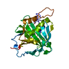
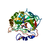
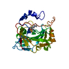
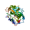
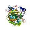
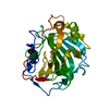
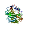
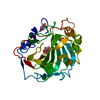
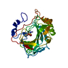
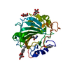
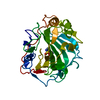
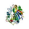
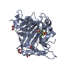
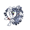
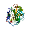
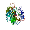
| Title | Crystal structure of human carbonic anhydrase XIII | ||||||
 Components Components | Carbonic anhydrase 13 | ||||||
 Keywords Keywords | METAL BINDING PROTEIN / Carbonic anhydrase / Lyase / Metal-binding | ||||||
| Function / homology |  Function and homology information Function and homology informationReversible hydration of carbon dioxide / carbonic anhydrase / carbonate dehydratase activity / myelin sheath / zinc ion binding / cytoplasm / cytosol Similarity search - Function | ||||||
| Biological species |  Homo sapiens (human) Homo sapiens (human) | ||||||
| Method |  X-RAY DIFFRACTION / SYNCHROTRON / MOLECULAR REPLACEMENT / Resolution: 1.55 Å X-RAY DIFFRACTION / SYNCHROTRON / MOLECULAR REPLACEMENT / Resolution: 1.55 Å | ||||||
 Authors Authors | Di Fiore, A. / De Simone, G. | ||||||
 Citation Citation | Journal: Proteins / Year: 2008 Title: Crystal structure of human carbonic anhydrase XIII and its complex with the inhibitor acetazolamide. Authors: Di Fiore, A. / Monti, S.M. / Hilvo, M. / Parkkila, S. / Romano, V. / Scaloni, A. / Pedone, C. / Scozzafava, A. / Supuran, C.T. / De Simone, G. #1: Journal: Proteins / Year: 1988Title: Refined structure of human carbonic anhydrase II at 2.0 A resolution. Authors: Eriksson, A.E. / Jones, T.A. / Liljas, A. | ||||||
| History |
|
- Structure visualization
| Structure viewer | Molecule: MolmilJmol/JSmol |
|---|
- Downloads & links
-Download
| PDBx/mmCIF format | 3d0n.cif.gz | 136.4 KB | Display | PDBx/mmCIF format |
|---|---|---|---|---|
| PDB format | pdb3d0n.ent.gz | 104 KB | Display | PDB format |
| PDBx/mmJSON format | 3d0n.json.gz | Tree view | PDBx/mmJSON format | |
| Others |  Other downloads Other downloads |
-Validation report
| Arichive directory | https://data.pdbj.org/pub/pdb/validation_reports/d0/3d0nftp://data.pdbj.org/pub/pdb/validation_reports/d0/3d0n | HTTPS FTP |
|---|
-Related structure data
| Related structure data |  3czvC 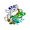 1ca2S S: Starting model for refinement C: citing same article ( |
|---|---|
| Similar structure data |


-Links
 PDBj
PDBj
- Assembly
| Deposited unit | 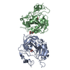
| ||||||||
|---|---|---|---|---|---|---|---|---|---|
| 1 | 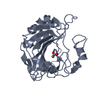
| ||||||||
| 2 | 
| ||||||||
| Unit cell |
|
-Components
| #1: Protein | Mass: 29626.252 Da / Num. of mol.: 2 Source method: isolated from a genetically manipulated source Source: (gene. exp.) Homo sapiens (human) / Gene: CA13 / Plasmid: pGEX-4T-1 / Production host:  #2: Chemical |   Mass: 65.409 Da / Num. of mol.: 2 / Source method: obtained synthetically / Formula: Zn Mass: 65.409 Da / Num. of mol.: 2 / Source method: obtained synthetically / Formula: Zn#3: Chemical |   Mass: 59.044 Da / Num. of mol.: 2 / Source method: obtained synthetically / Formula: C2H3O2 Mass: 59.044 Da / Num. of mol.: 2 / Source method: obtained synthetically / Formula: C2H3O2#4: Chemical | ChemComp-GOL / |   Mass: 92.094 Da / Num. of mol.: 1 / Source method: obtained synthetically / Formula: C3H8O3 Mass: 92.094 Da / Num. of mol.: 1 / Source method: obtained synthetically / Formula: C3H8O3#5: Water | ChemComp-HOH / |  Mass: 18.015 Da / Num. of mol.: 777 / Source method: isolated from a natural source / Formula: H2O Mass: 18.015 Da / Num. of mol.: 777 / Source method: isolated from a natural source / Formula: H2O |
|---|
-Experimental details
-Experiment
| Experiment | Method: X-RAY DIFFRACTION / Number of used crystals: 1 |
|---|
- Sample preparation
| Crystal | Density Matthews: 2.05 Å3/Da / Density % sol: 39.86 % |
|---|---|
| Crystal grow | Temperature: 293 K / Method: vapor diffusion, hanging drop / pH: 4.6 Details: 30% PEG 4000, 0.2 M ammonium acetate, 0.1 M sodium acetate pH 4.6, VAPOR DIFFUSION, HANGING DROP, temperature 293K |
-Data collection
| Diffraction | Mean temperature: 100 K |
|---|---|
| Diffraction source | Source: SYNCHROTRON / Site: ELETTRA  / Beamline: 5.2R / Wavelength: 0.999882 Å / Beamline: 5.2R / Wavelength: 0.999882 Å |
| Detector | Type: MAR CCD 165 mm / Detector: CCD / Date: Jun 1, 2007 |
| Radiation | Protocol: SINGLE WAVELENGTH / Monochromatic (M) / Laue (L): M / Scattering type: x-ray |
| Radiation wavelength | Wavelength: 0.999882 Å / Relative weight: 1 |
| Reflection | Resolution: 1.55→20 Å / Num. all: 68200 / Num. obs: 68200 / % possible obs: 98.1 % / Redundancy: 3.6 % / Rsym value: 0.058 / Net I/σ(I): 21.3 |
| Reflection shell | Resolution: 1.55→1.61 Å / Mean I/σ(I) obs: 3.8 / Num. unique all: 6269 / Rsym value: 0.258 / % possible all: 90.3 |
- Processing
| Software |
| |||||||||||||||||||||||||
|---|---|---|---|---|---|---|---|---|---|---|---|---|---|---|---|---|---|---|---|---|---|---|---|---|---|---|
| Refinement | Method to determine structure: MOLECULAR REPLACEMENT Starting model: PDB ENTRY 1CA2 Resolution: 1.55→20 Å / σ(F): 0 / Stereochemistry target values: Engh & Huber
| |||||||||||||||||||||||||
| Refinement step | Cycle: LAST / Resolution: 1.55→20 Å
| |||||||||||||||||||||||||
| Refine LS restraints |
|
